

Exposure of Teeth
Supporting optimal orthodontic alignment
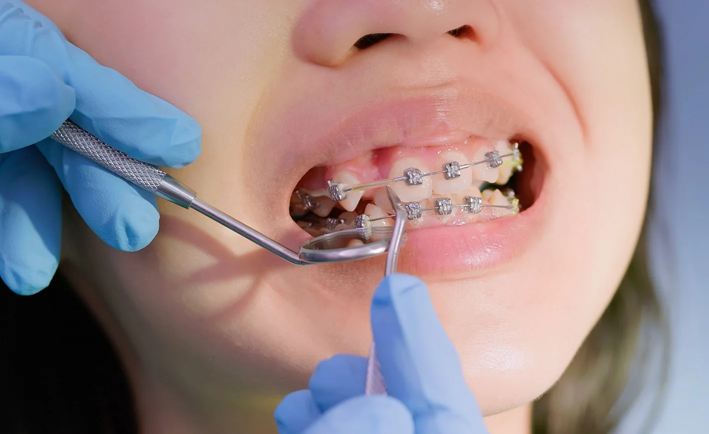
When a permanent tooth does not erupt naturally, surgical exposure can help guide it into the correct position as part of orthodontic treatment. This procedure is most commonly performed for impacted canine teeth and is carefully coordinated with the treating orthodontist.
Associate Professor Christian Freudlsperger and Mr Nigel Parr provide precise surgical care for impacted teeth, including complex cases involving unusual tooth positions, multiple impacted teeth, cysts or other obstructions. Treatment is individually planned to preserve the tooth and surrounding structures while supporting the best possible functional and aesthetic outcome.
UNDERSTANDING THE CONDITION
What is an impacted tooth?
An impacted tooth is a tooth that remains trapped beneath the gum or within the jawbone instead of erupting into its normal position. It may be blocked by another tooth, an over-retained baby tooth, insufficient space, dense bone, an abnormal eruption path or another condition within the jaw.
Which teeth commonly require exposure?
Canine Teeth: Upper canines are the second most commonly impacted teeth after wisdom teeth, affecting approximately 2-3% of the population. Canines are critical for proper bite function and facial aesthetics. They typically emerge around age 11-13, and their longer eruption path makes them more susceptible to impaction.
Other Teeth: Premolars, central and lateral incisors, second molars, and supernumerary (extra) teeth may also require exposure.
Why do teeth become impacted?
Several factors contribute to tooth impaction: insufficient space due to crowding, abnormal eruption path, early loss of baby teeth causing neighboring teeth to drift, retained baby teeth blocking eruption, extra teeth or cysts physically blocking the path, trauma disrupting tooth development, and genetic predisposition.
PROCEDURE & TREATMENT OPTIONS
Diagnosis & Treatment Planning
Comprehensive assessment includes clinical examination, advanced diagnostic imaging (panoramic X-rays, periapical X-rays, and 3D cone beam CT scans when necessary), and close collaboration with your orthodontist.
Imaging helps assess:
• The exact position and orientation of the impacted tooth
• Its relationship to neighbouring teeth and roots
• The amount of surrounding bone
• Root development and tooth condition
• The presence of cysts or other obstructions
• The most appropriate surgical approach
Close coordination with your orthodontist is essential. Orthodontic treatment is often started before surgery to create adequate space for the impacted tooth and prepare the dental arch for alignment.
The Exposure Procedure
Surgical exposure is usually performed as day treatment under local anaesthetic. Intravenous sedation may be offered for additional comfort or anxiety management, while general anaesthesia may be appropriate for younger patients, complex cases or when several procedures are required.
The surgical approach depends on the tooth’s position and the orthodontic treatment plan.
Soft Tissue Exposure (Open Eruption): When favorably positioned, a window of gum tissue is removed to expose the tooth crown, creating a clear path for natural or orthodontically-guided eruption.
Closed Eruption with Bonding: The most common approach involves creating a small flap in the gum tissue, carefully removing bone covering the crown, cleaning the exposed surface, bonding an orthodontic bracket or attachment with a small chain to the tooth, and repositioning the gum tissue with the chain extending through for orthodontic access. The orthodontist then applies gentle force to gradually guide the tooth into position over following months.
Additional Procedures: If impaction is caused by supernumerary teeth, cysts, or other obstructions, these are addressed during the same procedure. Bone grafting or soft tissue management may be performed when necessary.
Why Choose Oral & Facial Surgery, Remuera?
Specialist Surgical Expertise: Associate Professor Christian Freudlsperger and Mr Nigel Parr are specialist oral and maxillofacial surgeons experienced in the surgical management of impacted teeth, including complex and unusually positioned teeth.
Advanced Imaging and Precise Planning: Detailed radiographic assessment, including CBCT imaging where indicated, helps identify the safest surgical approach and protect neighbouring teeth and roots.
Close Orthodontic Collaboration: We work directly with orthodontists to coordinate timing, select the appropriate exposure technique and ensure the attachment is positioned to support controlled orthodontic movement.
Individualised, Age-Appropriate Care: Treatment and anaesthesia are tailored to the patient’s age, level of anxiety and procedural complexity, with local anaesthetic, IV sedation and general anaesthesia available where appropriate.
RISKS & RECOVERY
Understanding the Risks
Tooth exposure is safe and routine, but potential risks include mild to moderate pain and discomfort, swelling (resolving within 3-7 days), minor bleeding, bruising, temporary eating difficulties, infection (rare, treatable with antibiotics), damage to adjacent tooth roots (minimized through careful technique), loss of tooth vitality (occasionally requiring root canal treatment), ankylosis (rare fusion to bone preventing movement), and root resorption during orthodontic movement.
A/Prof Freudlsperger and Mr Nigel Parr will discuss all potential risks, benefits, and alternatives specific to your individual case in detail during your consultation, ensuring you have a complete understanding before proceeding with treatment.
Risks of Not Treating: Leaving impacted teeth untreated can cause damage to adjacent tooth roots, cyst formation, infection, compromised bite and aesthetics, tooth shifting, and more complex treatment requirements later.
What to Expect During Recovery
Mild discomfort, swelling and tenderness are expected for several days. Most patients can return to school, work and light daily activities within two to four days.
First 24-48 Hours: Some swelling and discomfort expected, peaking around day 2-3. Apply ice packs, take prescribed medication, maintain soft diet, avoid surgical area when brushing.
Days 3-7: Swelling subsides, discomfort decreases. Begin gentle salt water rinses. Many patients return to school or work within 2-4 days.
Week 2: Most swelling and discomfort resolved. Sutures removed if non-dissolvable (though dissolvable most common).
Ongoing Healing: Orthodontist typically waits 2-4 weeks before applying force to the bonded attachment.
Orthodontic Phase: Over 6-18 months, regular orthodontic visits guide the exposed tooth into position. Maintain excellent oral hygiene and wear prescribed appliances.
Post-Operative Care
Successful treatment depends on coordinated surgical and orthodontic follow-up. You will receive clear instructions regarding wound care, diet, oral hygiene and management of the orthodontic attachment.
Contact the practice if you experience increasing pain or swelling, persistent bleeding, signs of infection or detachment of the orthodontic chain.
Associate Professor Freudlsperger and Mr Parr work closely with your orthodontist throughout treatment to support safe healing and the successful alignment of the impacted tooth.
BOOK YOUR APPOINTMENT
Oral & Facial Surgery Auckland
If you or your child has been diagnosed with an impacted tooth, or if you suspect a tooth has failed to erupt properly, A/Prof Christian Freudlsperger can help. With internationally recognized expertise and a collaborative approach to treatment, he works closely with orthodontists to achieve excellent outcomes.
Book your Appointment
Lorem ipsum dolor sit amet, consectetur adipiscing elit, sed do eiusmod tempor incididunt ut labore et dolore magna aliqua. Dui faucibus in ornare quam. Lectus urna duis convallis convallis tellus id interdum velit laoreet.