

Skin Surgery
Precise treatment for facial skin lesions and skin cancer
Skin lesions on the face require careful assessment because treatment must address the abnormal tissue while preserving facial function, contour and appearance. This is especially important around the lips, eyelids, nose, cheeks, ears and jawline, where even a small defect can affect movement, symmetry or facial expression.
Associate Professor Christian Freudlsperger provides specialist assessment, biopsy, excision and reconstruction of benign and malignant facial skin lesions. His background in oral and maxillofacial, oncological and reconstructive surgery supports careful treatment planning for lesions in anatomically and cosmetically sensitive areas.
Treatment may range from a minor procedure under local anaesthetic to local flap reconstruction following removal of a larger skin cancer or after Mohs surgery. Excised tissue is commonly sent for histopathological examination to confirm the diagnosis and assess whether the lesion has been completely removed.
UNDERSTANDING THE PROCEDURE
Types of Benign Skin Lesions
Moles
Moles, or naevi, may be flat or raised and vary in colour. Most are harmless, but a new or changing pigmented lesion should be assessed to exclude melanoma or another skin cancer.
Changes that warrant review include:
Increasing size
Irregular shape or border
Multiple or changing colours
Bleeding, crusting or ulceration
Persistent itching or tenderness
A lesion that looks different from the patient’s other moles
Skin Tags
Skin tags are small, soft growths commonly found on the eyelids, neck or other areas of friction. Removal may be considered if they become irritated, bleed or cause concern.
Seborrhoeic Keratoses
These are common benign growths that may appear waxy, scaly or “stuck on”. They usually require no treatment unless they are irritated, diagnostically uncertain or cosmetically troublesome.
Skin Cysts
Epidermoid and similar cysts form beneath the skin and may become swollen, infected or painful.
To reduce recurrence, the cyst lining should generally be removed completely rather than simply drained.
Lipomas
Lipomas are soft, mobile collections of fatty tissue beneath the skin. Removal may be considered when they enlarge, cause discomfort or affect facial contour.
Facial Skin Cancer
Basal Cell Carcinoma
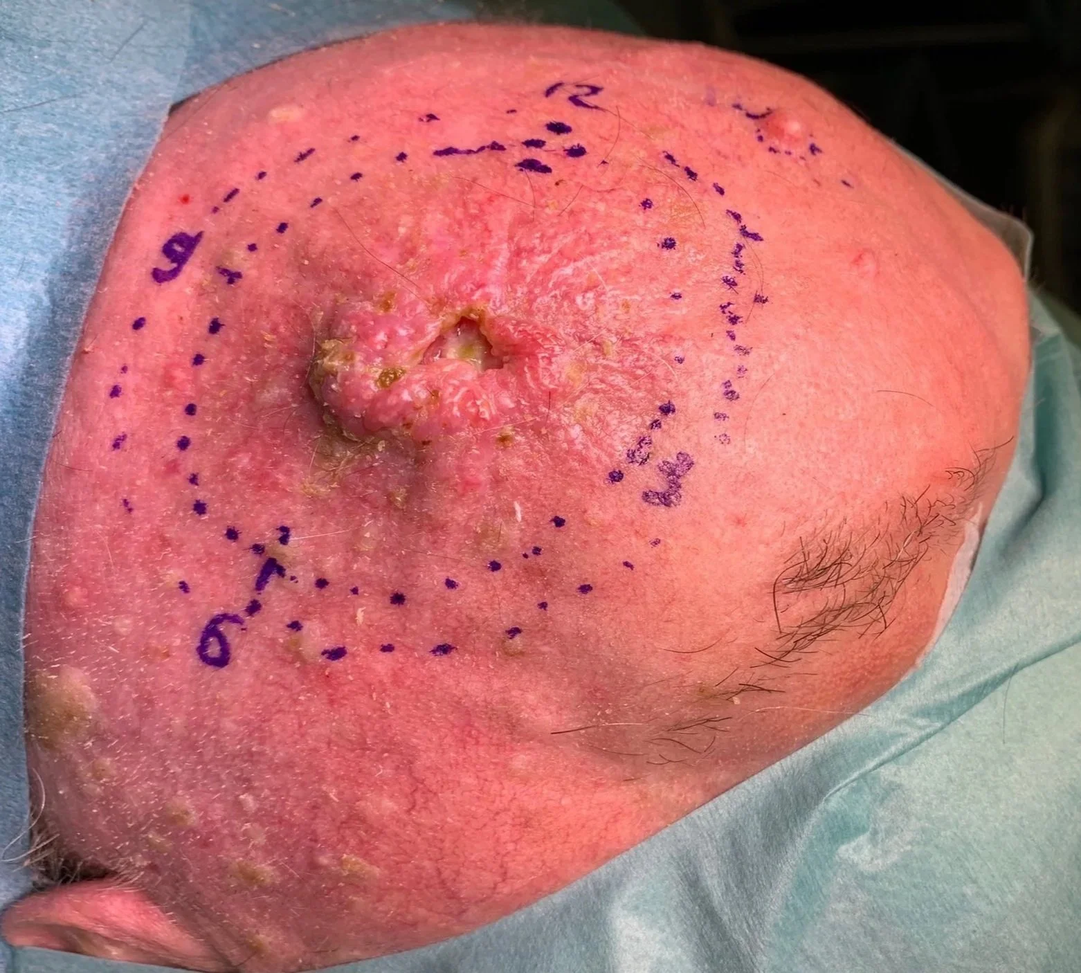
Basal cell carcinoma is the most common skin cancer. It commonly develops on sun-exposed facial skin and may appear as:
A pearly or translucent lump
A non-healing sore
A pink patch
A lesion that repeatedly bleeds or crusts
A scar-like area without previous injury
Basal cell carcinoma rarely spreads to distant areas, but it can invade nearby tissue and cause significant local damage if left untreated.
Cutaneous Squamous Cell Carcinoma
Cutaneous squamous cell carcinoma may appear as:
A rough or scaly patch
A firm raised growth
A rapidly growing lump
A persistent ulcer
A lesion that bleeds or becomes painful
Some squamous cell carcinomas have a risk of spreading to lymph nodes, particularly when they are large, recurrent, deeply invasive or located in higher-risk areas such as the ear or lip.
Melanoma
Melanoma requires prompt specialist assessment. Suspicious pigmented lesions are generally referred through an appropriate dermatology, melanoma or multidisciplinary pathway.
When should a Skin Lesion be assessed?
Seek medical review for:
A sore that has not healed within several weeks
A new or enlarging facial lump
A changing mole
Persistent bleeding or crusting
An ulcerated skin lesion
A rapidly growing lesion
A painful or numb area
A recurrent lesion after previous treatment
A lesion close to the eye, lip, nose or ear
A new lump in the neck or near the parotid gland
PROCEDURE & TREATMENT OPTIONS
Assessment and Diagnosis
Your consultation may include:
Review of how long the lesion has been present
Assessment of recent changes or symptoms
Clinical examination of the lesion and surrounding skin
Examination of nearby facial nerves and lymph nodes where appropriate
Review of previous biopsies or treatment
Clinical photography
Biopsy or complete excision
Imaging in selected larger or more complex cases
Some lesions can be diagnosed clinically. Others require tissue sampling because benign and malignant skin lesions may occasionally look similar.
Biopsy Procedures
Incisional Biopsy
A representative part of a larger lesion is removed for diagnosis before definitive treatment is planned.
Excisional Biopsy
The entire lesion is removed, usually with a small margin of surrounding tissue. This may provide both diagnosis and treatment for smaller lesions.
Punch Biopsy
A circular instrument removes a small full-thickness sample. This can be useful when only a diagnostic specimen is required.
Shave Biopsy or Shave Excision
A raised superficial lesion is removed at or just below the skin surface. This is suitable only for selected lesions and may not be appropriate where full-depth assessment or margin evaluation is required.
Surgical Excision
Surgical excision removes the lesion together with an appropriate margin of surrounding tissue.
The required margin depends on:
The diagnosis
Tumour subtype
Lesion size and depth
Anatomical location
Previous treatment
Histopathological risk features
The specimen is sent for histopathological examination. Further treatment may be recommended if the diagnosis is unexpected or the margins are incomplete.
Simple Wound Closure
Smaller defects may be closed directly with carefully positioned sutures.
For facial surgery, the incision is planned with attention to:
Natural skin creases
Relaxed skin tension lines
Facial aesthetic units
Eyelid and lip function
Avoidance of distortion of nearby structures
Minimisation of tension across the wound
Layered closure may be used to support the deeper tissues and improve scar quality.
Local Flap Reconstruction
When a defect cannot be closed directly without distortion or excessive tension, nearby skin may be repositioned as a local flap.
Local flaps use tissue with a similar colour, thickness and texture to the removed skin and may provide a better result than a graft in selected facial areas.
They are commonly used around the:
Nose
Eyelids
Lips
Cheeks
Ears
Forehead
Jawline
The reconstruction is selected according to the size, shape and location of the defect and the need to preserve movement and facial symmetry.
Skin Grafting
A skin graft may be used when local tissue rearrangement is unsuitable.
Skin is taken from another site and placed over the defect. The colour and contour may differ from the surrounding facial skin, but grafting can provide reliable coverage in selected cases.
Reconstruction after Mohs Surgery
Mohs micrographic surgery is commonly performed by specially trained dermatologists for selected high-risk or anatomically sensitive skin cancers.
After the cancer has been removed and the margins confirmed, reconstruction may involve:
Direct closure
Local flap reconstruction
Skin grafting
Staged reconstruction
Repair of the lip, eyelid, nose or ear
Associate Professor Freudlsperger can provide reconstruction following Mohs or dermatological excision in selected cases, with the aim of preserving function and achieving the best possible facial contour.
Scar Revision
Scar revision may be considered for scars that are:
Widened
Raised or thickened
Contracted
Depressed
Poorly aligned with facial skin lines
Distorting the lip, eyelid or another structure
No procedure can remove a scar completely, but revision may improve its position, width or contour.
Why Choose A/Prof Christian Freudlsperger?
Specialist Facial Surgical Expertise: Associate Professor Christian Freudlsperger has more than 20 years of experience in oral and maxillofacial, oncological and reconstructive surgery, with detailed knowledge of facial anatomy and function.
Careful Cancer and Pathology Assessment: Biopsy, excision and histopathological examination are used to establish an accurate diagnosis and guide any further treatment.
Reconstructive Capability: Treatment ranges from simple excision and layered closure to local flaps, skin grafts and reconstruction following Mohs or cancer surgery.
Function and Appearance Considered Together: Surgical planning aims to remove the lesion safely while preserving facial movement, symmetry and natural anatomical landmarks.
Associate Professor Freudlsperger’s research has also examined advanced optical imaging and artificial-intelligence-assisted analysis of basal cell carcinoma, oral squamous cell carcinoma, oral leukoplakia and normal oral tissues. This academic work reflects an ongoing interest in improving tissue diagnosis and distinguishing abnormal from healthy tissue.
RISKS
Understanding the Risks of Chin Surgery
While lesion removal is generally straightforward, there are potential risks to any surgery.
A/Prof Freudlsperger will discuss all potential risks, benefits, and realistic expectations about scarring specific to your lesion location and type during your consultation. Understanding that scarring is inevitable and its appearance cannot be guaranteed is essential before proceeding.
Common Temporary Effects:
Some discomfort at removal site
Swelling and bruising (particularly for larger excisions)
Incision or wound requiring care during healing
Temporary redness and firmness
Activity restrictions during initial healing
Potential Complications:
Scarring: This is inevitable with surgical removal. All techniques that remove lesions create scars, though appearance varies significantly based on location, size, skin type, healing characteristics, and technique. Scars on the face generally heal well but are visible on close inspection. Body scars may be more prominent. Thick, raised (hypertrophic) or widened scars can develop despite optimal technique.
Scar appearance not meeting expectations: While surgeons aim for optimal scars, individual healing determines final appearance and cannot be entirely predicted
Recurrence: Incomplete removal can result in lesion regrowth, particularly with shave techniques or if cyst wall not completely removed
Infection: Uncommon but can compromise healing and worsen scarring
Bleeding or hematoma: Blood collection at site requiring drainage
Wound dehiscence: Incision separation during healing, may require additional treatment
Nerve damage: Rarely, small sensory nerves can be affected, causing numbness or altered sensation
Pigmentation changes: Hyper- or hypopigmentation can occur, particularly in darker skin types
Keloid formation: In predisposed individuals, particularly over chest, shoulders, and earlobes
Need for scar revision: Some scars may benefit from revision procedures, though this cannot guarantee perfect results
Pathology findings: Rarely, lesions thought benign prove atypical or concerning on pathology, requiring additional treatment
Specific Risk Factors:
Family history of abnormal scarring (keloids, hypertrophic scars)
Darker skin types have increased risk of pigmentation changes
Certain body locations (chest, shoulders, back) have higher risk of thick scarring
Smoking impairs healing and increases complications
Location over tension areas or mobile areas may result in wider scars
Realistic Expectations:
All removal techniques create scars
You are trading a lesion for a scar
Scar will be permanent and visible on close inspection
Scar appearance improves over 12-18 months but won't disappear
Individual healing characteristics significantly affect scar quality
Perfect, invisible scars are not possible
Some locations scar more favorably than others
Scars on the face generally heal better than body scars
Width of scar depends on tension and healing, not just surgeon skill
Some patients may be disappointed that scar is more visible than expected
Makeup can usually camouflage facial scars once healed
YOUR RECOVERY
What to Expect During Recovery
Most small facial skin procedures are performed under local anaesthetic as outpatient treatment.
First 24–48 Hours
Mild discomfort, swelling and bruising are common. The dressing should be kept clean and dry according to the postoperative instructions.
A small amount of blood staining may be normal, but persistent bleeding should be reported.
First Week
Facial sutures are often removed after approximately five to seven days, depending on the location and wound tension.
Sutures in other areas may remain longer.
Patients should avoid:
Strenuous activity
Stretching the wound
Swimming
Makeup over the incision
Picking at scabs or sutures
Smoking and vaping
Following Weeks
The scar may initially look pink, red or slightly raised. This does not necessarily indicate a poor result.
Once the wound has healed, scar management may include:
Silicone gel or silicone sheets
Gentle scar massage
Moisturising
Sun protection
Further review if the scar becomes thickened or symptomatic
Scar Maturation
Scars commonly continue to soften and fade for 12–18 months.
Strict sun protection is important because ultraviolet exposure can cause prolonged redness or pigmentation.
Frequently Asked Questions
1. How do I know whether a facial skin lesion is cancer?
It is not always possible to determine this from appearance alone.
Warning signs include growth, bleeding, ulceration, colour change, irregular borders or failure to heal. Clinical assessment and biopsy or excision may be needed to establish the diagnosis.
2. Does every mole need to be removed?
No. Most moles are benign and can be observed.
Removal may be recommended if the mole is changing, symptomatic, diagnostically uncertain or causing significant concern. Suspicious pigmented lesions may require dermatological or melanoma assessment.
3. Will skin cancer surgery leave a scar?
Yes. Any procedure that cuts the skin leaves a permanent scar.
The incision and reconstruction are carefully planned to place the scar within natural facial lines where possible and reduce tension or distortion. The final appearance continues to improve over many months.
4. Is all removed tissue sent to the laboratory?
Lesions removed for diagnostic or cancer-related reasons are generally sent for histopathological examination.
The pathologist confirms the diagnosis and, where relevant, assesses whether the lesion has been completely removed.
5. What happens if the skin cancer has not been completely removed?
Further excision may be recommended to obtain clear margins.
The next step depends on the cancer type, location, remaining margin and individual risk features. In some cases, referral for Mohs surgery, multidisciplinary review or another treatment may be appropriate.
Selected Research & Publications:
Shavlokhova V, Vollmer M, Gholam P, Saravi B, Vollmer A, Hoffmann J, Engel M, Freudlsperger C. Deep learning on basal cell carcinoma in vivo reflectance confocal microscopy data. Journal of Personalized Medicine. 2022;12(9):1471.
Shavlokhova V, Flechtenmacher C, Sandhu S, Vollmer M, Vollmer A, Saravi B, Engel M, Ristow O, Hoffmann J, Freudlsperger C. Ex vivo fluorescent confocal microscopy images of oral mucosa: tissue atlas and evaluation of the learning curve. Journal of Biophotonics. 2022;15(2):e202100225.
Shavlokhova V, Sandhu S, Flechtenmacher C, Koveshazi I, Neumeier F, Padrón-Laso V, Jonke Ž, Saravi B, Vollmer M, Vollmer A, Hoffmann J, Engel M, Ristow O, Freudlsperger C. Deep learning on oral squamous cell carcinoma ex vivo fluorescent confocal microscopy data: a feasibility study. Journal of Clinical Medicine. 2021;10(22):5326.
Shavlokhova V, Flechtenmacher C, Sandhu S, Vollmer M, Vollmer A, Pilz M, Hoffmann J, Ristow O, Engel M, Freudlsperger C. Feasibility and implementation of ex vivo fluorescence confocal microscopy for diagnosis of oral leukoplakia: preliminary study. Diagnostics. 2021;11(6):951.
BOOK YOUR APPOINTMENT
Schedule Your Lesion Removal Consultation
Please arrange an assessment if you have a new, changing, bleeding or non-healing facial skin lesion, or require reconstruction following skin-cancer or Mohs surgery.
Referrals are welcome from dermatologists, GPs, plastic surgeons, Mohs surgeons, ophthalmologists, ENT surgeons, dentists and other medical specialists.