

Facial Trauma
Specialist treatment for facial fractures, jaw injuries and post-traumatic deformity
Facial trauma can affect much more than appearance. Injuries to the jaws, cheekbones, eye sockets and midface may alter the bite, breathing, vision, facial sensation, speech, chewing and jaw movement.
Prompt specialist assessment helps identify fractures that require treatment and reduces the risk of persistent problems such as malocclusion, facial asymmetry, restricted mouth opening, double vision or chronic discomfort.
Associate Professor Christian Freudlsperger provides specialist oral and maxillofacial surgical care for acute facial injuries and secondary reconstruction of fractures that have healed in an unfavourable position. He has more than 20 years of international surgical experience and is Service Clinical Director of Auckland’s regional Oral and Maxillofacial Surgery service. The regional service manages fractures of the jaws, cheekbones and eye sockets at Middlemore and Auckland City Hospitals.
Advanced CT imaging, virtual surgical planning and modern reconstructive techniques are used where appropriate to restore facial structure, function and symmetry.
UNDERSTANDING THE CONDITION
Types of Facial Injuries
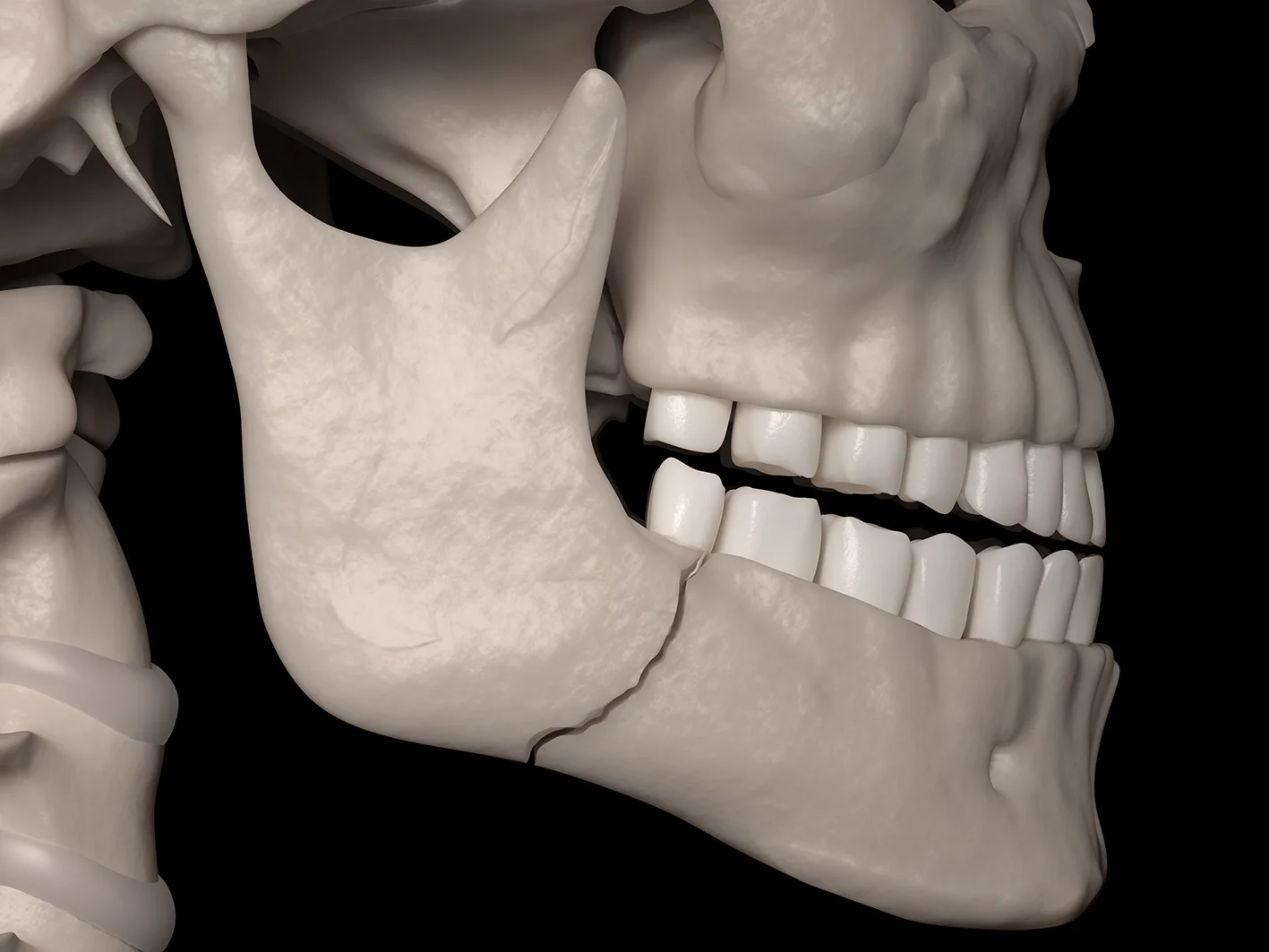
1. Mandibular Fractures
A mandibular fracture affects the lower jaw and may involve:
The tooth-bearing body of the jaw
The angle near the back teeth
The vertical ramus
The mandibular condyle near the jaw joint
The chin or symphysis
More than one region of the mandible
Possible consequences include an altered bite, pain when chewing, restricted mouth opening, numbness of the lower lip and instability of the jaw.
2. Maxillary and Midface Fractures
Fractures of the upper jaw and central face may affect the relationship between the upper and lower teeth, facial height, nasal airway and support of the cheeks and eyes.
Complex midface injuries may involve several bones and require careful restoration of facial width, height and projection.
3. Zygomatic or Cheekbone Fractures
The zygoma forms the prominence of the cheek and part of the eye socket.
A displaced cheekbone fracture may cause:
Flattening of the cheek
Facial asymmetry
Numbness of the cheek, upper lip or side of the nose
Difficulty opening the mouth
Double vision
Changes in eye position
4. Orbital Fractures
Orbital fractures involve the bones surrounding the eye.
They may cause:
Double vision
Restricted eye movement
Blurred or reduced vision
A sunken or displaced eye
Numbness of the cheek and upper lip
Swelling around the eye
Any deterioration in vision, severe eye pain or limitation of eye movement requires urgent medical assessment.
5. Nasal and Naso-Orbito-Ethmoidal Injuries
Nasal fractures may cause deformity, bleeding or difficulty breathing through the nose.
More complex central facial fractures can affect the nasal bridge, tear ducts, inner eye support and attachment of the eyelids.
6. Dentoalveolar Trauma
Dentoalveolar injuries involve the teeth and the bone supporting them.
They may include:
Chipped or fractured teeth
Displaced teeth
Avulsed or knocked-out teeth
Root fractures
Fracture of the tooth-bearing jawbone
Injury to developing permanent teeth in children
These injuries often require coordinated care between the surgeon, dentist, endodontist and orthodontist.
7. Facial Soft-Tissue Injuries
Facial cuts may involve the lips, eyelids, nose, cheek, nerves or salivary ducts.
Careful repair aims to restore normal tissue alignment, preserve function and minimise scarring, particularly around important landmarks such as the lip margin and eyelids.Common Causes
Facial trauma results from motor vehicle accidents, sports injuries (particularly contact sports), falls (especially in elderly or intoxicated individuals), physical assaults, workplace accidents, and bicycle or pedestrian accidents.
CAUSES & SYMPTOMS
Common causes of Facial Trauma
Facial trauma may result from:
Motor vehicle, bicycle or pedestrian accidents
Sports injuries
Falls
Physical assault
Workplace accidents
Animal-related injuries
Other accidental impacts
Signs & Symptoms
Specialist assessment may be required for:
Facial swelling or significant bruising
Jaw pain or instability
A change in the way the teeth meet
Difficulty opening or closing the mouth
Loose, displaced or damaged teeth
Facial numbness
Flattening or asymmetry of the cheek
Double vision or restricted eye movement
A sunken or displaced eye
Nasal obstruction or deformity
Persistent bleeding
A wound involving the lip, eyelid or another important facial structure
PROCEDURE & TREATMENT OPTIONS
Emergency Assessment & Initial Management
Facial trauma requires prompt evaluation to assess injury severity and plan appropriate treatment. Initial assessment includes airway evaluation (ensuring breathing isn't compromised), bleeding control, clinical examination of facial bones and soft tissues, neurological assessment (nerve function, vision), and advanced imaging (CT scans for bone injuries, X-rays for dental injuries).
Many facial injuries are initially assessed in hospital emergency departments, where A/Prof Freudlsperger provides consultant services.
Treatment Approaches
Soft Tissue Repair: Meticulous layered closure of facial lacerations using fine sutures, careful alignment of tissue planes and landmarks (vermillion border of lips, eyebrows), microsurgical nerve repair when necessary, and primary closure within 24 hours optimal for best outcomes and minimizing scarring.
Dentoalveolar Treatment: Repositioning and stabilization of displaced teeth, splinting of mobile teeth, management of alveolar fractures, and tooth replantation if avulsed teeth brought to hospital in appropriate medium within short timeframe.
Mandible Fracture Treatment: Non-displaced, stable fractures may be managed with soft diet and careful monitoring. Displaced or unstable fractures require open reduction and internal fixation—surgical exposure of fracture, precise realignment, and stabilization with titanium plates and screws. Some fractures can be managed with closed reduction and maxillomandibular fixation (wiring jaws together temporarily).
Midface Fracture Treatment: Zygoma (cheekbone) fractures often require open reduction and fixation to restore facial projection and symmetry. Orbital fractures may require reconstruction with plates or grafts to prevent eye complications. Maxillary fractures require reduction and fixation, sometimes with temporary maxillomandibular fixation. Nasal fractures are reduced and stabilized as appropriate.
Complex Panfacial Trauma: Severe injuries involving multiple facial bones require staged reconstruction in a specific sequence to restore facial height, width, and projection. These complex cases benefit significantly from A/Prof Freudlsperger's extensive trauma experience.
Timing of Treatment
Some injuries require immediate treatment (active bleeding, airway compromise, globe injury), while others can be addressed semi-urgently within days (most facial fractures, many soft tissue injuries). Delayed treatment may be necessary when significant swelling obscures anatomy or patient has other life-threatening injuries requiring stabilization first.
SECONDARY FACIAL TRAUMA RECONSTRUCTION
When a Facial Fracture Heals in the Wrong Position
A fracture that heals in an incorrect position is called a malunion. A fracture that fails to unite properly is called a non-union.
Patients may seek assessment weeks, months or years after an injury because of:
Facial asymmetry
Flattening or widening of the cheek
A displaced jaw or chin
Persistent bite problems
Restricted mouth opening
Difficulty chewing
Double vision
A sunken eye
Nasal obstruction
Persistent numbness
Painful or prominent plates
Contour irregularity following previous surgery
Secondary reconstruction is more complex than treating a recent fracture because the bones have already healed and scar tissue may have altered the normal anatomy.
Assessment of an Old Facial Injury
Planning may include:
Review of the original injury and previous operations
Clinical photographs
Examination of facial symmetry and contour
Dental and bite analysis
Assessment of eye position and movement
CT imaging with three-dimensional reconstruction
Comparison with the unaffected side
Digital dental and facial scans
Review of current plates or implants
Ophthalmology or orthodontic assessment where required
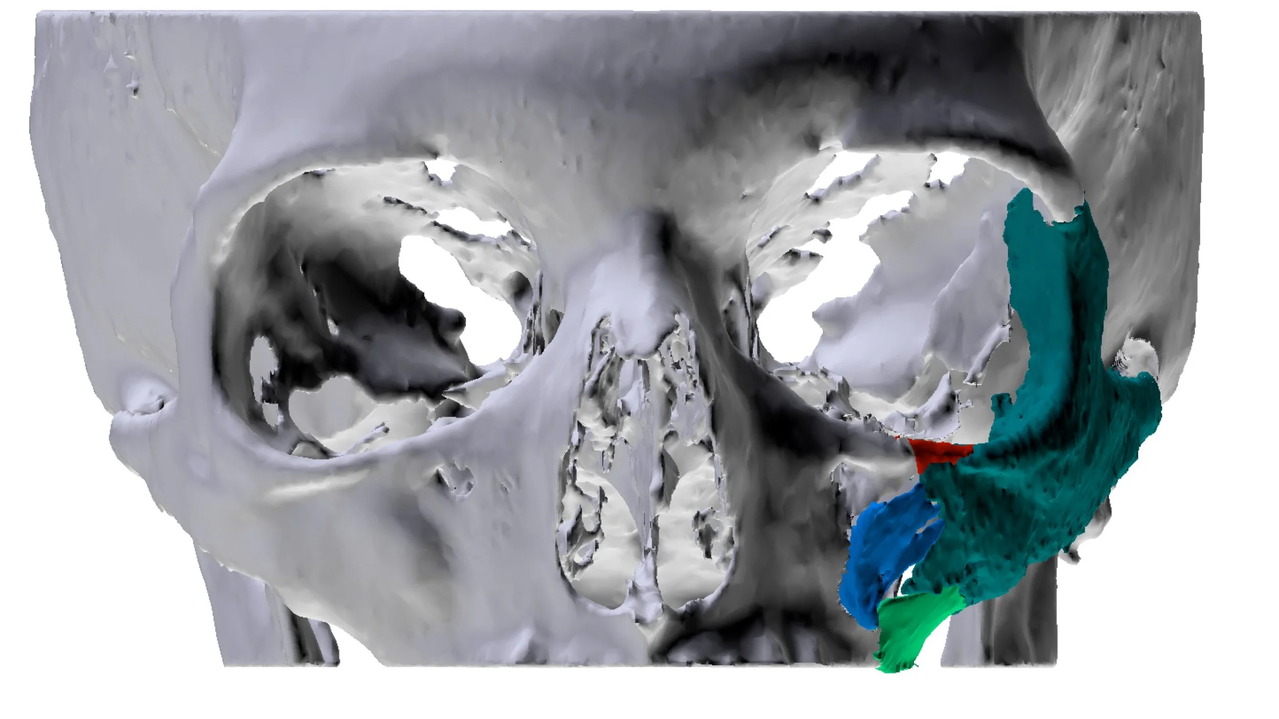
Virtual surgical planning for secondary reconstruction
Virtual surgical planning is particularly valuable for fractures that have healed in a displaced position.
It may be used to:
Identify the original fracture pattern
Recreate the expected pre-injury anatomy
Mirror the unaffected side of the face
Plan corrective bone cuts
Reposition the jaws or cheekbones digitally
Restore orbital volume and eye support
Design cutting or positioning guides
Plan patient-specific implants
Coordinate correction of the bite
The objective is to restore facial anatomy as closely and safely as possible while addressing the patient’s main functional and appearance concerns.
Secondary Reconstructive Procedures
Treatment may involve:
Corrective osteotomy and repositioning of a malunited fracture
Revision fixation with plates and screws
Bone grafting
Patient-specific implants
Orbital wall reconstruction
Correction of cheekbone projection
Corrective jaw surgery for post-traumatic malocclusion
Scar revision
Removal or replacement of symptomatic hardware
Dental, orthodontic or implant rehabilitation
Treatment may be completed in one operation or staged over time depending on the extent of the deformity.
Why Choose A/Prof Christian Freudlsperger?
Specialist Facial Trauma Experience: Associate Professor Christian Freudlsperger has extensive international experience in the treatment of mandibular, maxillary, cheekbone, orbital and complex facial fractures.
Current Regional Trauma Practice: As Service Clinical Director of Auckland’s regional Oral and Maxillofacial Surgery service, he works within a service that undertakes facial fracture surgery at Middlemore and Auckland City Hospitals.
Advanced Secondary Reconstruction: Particular expertise in virtual surgical planning and complex facial reconstruction supports correction of malunited fractures, post-traumatic asymmetry, orbital deformity and persistent bite problems.
Restoration of Function and Appearance: Treatment considers the bite, jaw movement, vision, airway, facial sensation, symmetry and long-term dental rehabilitation rather than focusing on the fracture alone.
ACC FUNDING
ACC & Facial Trauma Treatment
In New Zealand, facial injuries caused by an accident may be covered by ACC. ACC’s no-fault accident scheme covers eligible accidental injuries, and it may contribute to medical, surgical and rehabilitation costs when a claim is accepted. Patients may still need to pay part of the treatment cost, depending on the provider, procedure and applicable ACC funding arrangements.
ACC funding may potentially apply to:
Assessment of an accidental facial injury
Treatment of jaw or facial fractures
Dental trauma
Removal of symptomatic fixation hardware
Further treatment for persistent effects of a covered injury
Secondary reconstruction of an accepted injury
Rehabilitation and selected support services
Coverage and prior approval are assessed individually. Please bring your ACC claim number, original accident details, previous clinical records and imaging to the consultation where available.
RISK & RECOVERY
Understanding the Risks
Facial trauma treatment carries potential risks depending on injury severity: infection (managed with antibiotics and wound care), scarring (minimized through meticulous technique but inevitable with lacerations), malunion or nonunion of fractures (improper or failed bone healing), malocclusion (bite problems requiring orthodontic correction), persistent numbness from nerve injury (often improves over months but may be permanent), diplopia (double vision from orbital injuries), and need for secondary procedures (scar revision, bone contouring, orthodontics).
A/Prof Freudlsperger will discuss all potential risks, benefits, and alternatives specific to your individual case in detail during your consultation, ensuring you have a complete understanding before proceeding with treatment.
What to Expect During Recovery
Acute Phase (First Week): Significant swelling and bruising (peaking at 48-72 hours), pain managed with prescribed medication, soft or liquid diet depending on injury, activity restriction, and possible jaw fixation limiting mouth opening.
Weeks 2-6: Swelling and bruising gradually resolve, sutures removed (facial sutures typically 5-7 days, intraoral 10-14 days), gradual return to normal diet as healing permits, return to light activities, and avoid contact sports or activities risking re-injury.
Months 2-6: Continued improvement in swelling, numbness, and function. Bone healing completes over 6-8 weeks. Physical therapy for jaw movement if needed. Scar maturation continues for 12-18 months.
Long-Term: Most patients achieve excellent functional recovery and acceptable aesthetics. Secondary procedures occasionally needed for scar revision, bone contouring, or orthodontic treatment.
Post-Operative Care
Success depends on following instructions: maintain excellent oral hygiene (gentle initially), follow dietary restrictions as directed, take antibiotics if prescribed, manage pain with prescribed medication, avoid smoking (severely impairs healing), avoid contact sports or high-risk activities for 6-12 weeks, attend all follow-up appointments, and report concerning symptoms immediately (fever, increasing pain, visual changes, excessive swelling).
Early, expert treatment of facial trauma provides the best opportunity for optimal healing and restoration of your appearance and function.
Frequently Asked Questions
1. How do I know whether my jaw is fractured?
Possible signs include jaw pain, swelling, an altered bite, difficulty opening the mouth, numbness of the lower lip, loose teeth or movement of the jaw at an abnormal point.
A clinical examination and appropriate imaging are required to confirm the diagnosis.
2. Does every mandibular fracture need surgery?
No. Stable and minimally displaced fractures may sometimes be managed with a soft diet, elastics or close observation.
Surgery is more likely when the fracture is displaced, unstable, open, associated with an altered bite or unlikely to heal predictably without fixation.
3. Will my jaw be wired shut after fracture surgery?
Not usually for a prolonged period. Modern titanium plates and screws often provide stable fixation while allowing earlier movement.
Temporary elastics or short-term fixation may still be used to guide the bite, depending on the fracture.
4. How long does a broken jaw take to heal?
Initial bone healing commonly takes around six weeks, although recovery varies with the fracture pattern, age, health, smoking status and treatment.
A soft diet and protection from further injury are generally required during early healing.
5. Can a facial fracture that healed incorrectly be repaired later?
Often, yes. Secondary facial trauma reconstruction can improve malocclusion, asymmetry, cheek flattening, orbital deformity, double vision and other persistent problems.
Treatment may require corrective bone cuts, repositioning, bone grafting or patient-specific implants. CT imaging and virtual surgical planning help determine what can realistically be achieved.Virtual Surgical Planning for Secondary Reconstruction.
BOOK YOUR APPOINTMENT
Experienced Emergency Facial Trauma Care
Please seek urgent hospital care for any recent facial injury associated with breathing difficulty, heavy bleeding, reduced vision, loss of consciousness or severe deformity.
For non-emergency assessment, persistent symptoms or secondary reconstruction of an old facial injury, please contact Oral & Facial Surgery to arrange a consultation with Associate Professor Christian Freudlsperger.
Referrals are welcome from emergency physicians, GPs, dentists, orthodontists, ophthalmologists, ENT surgeons, plastic surgeons and other medical specialists.