

Salivary Gland Surgery
Specialist assessment and treatment of salivary stones, cysts and tumours
The salivary glands can be affected by stones, recurrent infections, cysts, persistent swelling and benign or malignant tumours. Symptoms may include pain or swelling during meals, a lump near the ear or beneath the jaw, dry mouth, repeated infections or reduced facial movement.
Many salivary gland conditions are benign. However, any persistent or unexplained lump in the parotid region, beneath the jaw, in the neck or inside the mouth should be assessed to establish the cause and determine whether treatment is required.
Associate Professor Christian Freudlsperger provides specialist assessment and surgical management of conditions affecting the parotid, submandibular, sublingual and minor salivary glands. He has more than 20 years of international experience in oral and maxillofacial and head and neck surgery and is Service Clinical Director of Auckland’s regional Oral and Maxillofacial Surgery service, which includes the management of salivary gland disease
UNDERSTANDING THE ANATOMY
What Are the Salivary Glands?
The salivary glands produce saliva, which moistens the mouth, supports swallowing, protects the teeth and oral tissues and begins the digestion of food.
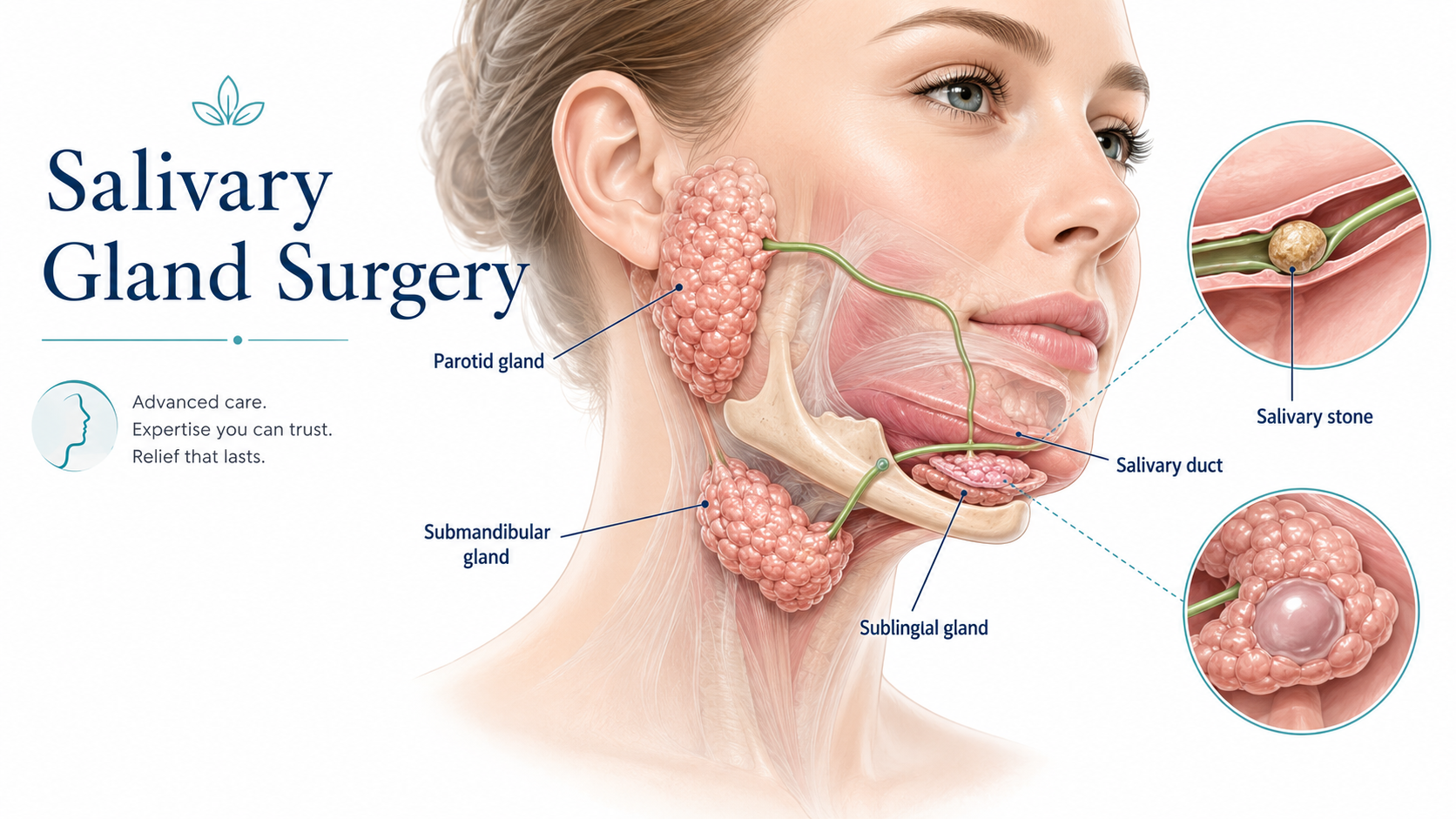
There are three pairs of major salivary glands:
Parotid Glands
The parotid glands are located in front of and below each ear. Saliva passes from each gland through a duct that opens inside the cheek.
The facial nerve passes through the parotid gland and divides into branches that control facial movement. Protecting and monitoring facial nerve function is therefore a central consideration during parotid surgery.
Submandibular Glands
The submandibular glands sit beneath the lower jaw. Their ducts travel through the floor of the mouth and open beneath the tongue.
Stones occur frequently in the submandibular duct and may cause pain or swelling during meals.
Sublingual Glands
The sublingual glands are located beneath the tongue. Blockage or leakage of saliva from this region can contribute to the development of a ranula.
Minor Salivary Glands
Hundreds of small salivary glands are distributed throughout the lips, cheeks, palate, tongue and throat. They may develop cysts, inflammatory conditions or tumours.
UNDERSTANDING THE ANATOMY
Conditions Affecting the Salivary Glands
Salivary Stones
Salivary stones, also called sialolithiasis, can obstruct the flow of saliva through a gland or duct.
Symptoms may include:
Pain or swelling during meals
Recurrent swelling beneath the jaw or inside the mouth
Reduced saliva flow
Tenderness of the gland
An unpleasant taste or discharge
Infection around the obstructed duct
Meal-related pain and swelling are characteristic because saliva production increases when eating but cannot pass freely through the blocked duct.
Sialadenitis
Sialadenitis is inflammation or infection of a salivary gland.
It may be associated with:
A salivary stone or duct narrowing
Dehydration or reduced saliva flow
Bacterial or viral infection
Autoimmune disease
Previous radiotherapy
Recurrent obstruction
Symptoms can include pain, swelling, redness, fever or discharge from the duct.
Mucoceles and Ranulas
A mucocele is a saliva-filled swelling that usually develops after injury or blockage of a minor salivary gland duct, commonly inside the lower lip.
A ranula develops in the floor of the mouth, usually in association with the sublingual gland. Larger ranulas may extend into the neck and are sometimes called plunging ranulas.
Benign Salivary Gland Tumours
Many salivary gland tumours are benign. Pleomorphic adenoma is a common benign parotid tumour and often presents as a slow-growing, painless lump.
Although benign, some tumours continue to enlarge and may become more difficult to remove safely over time. Pleomorphic adenomas also carry a long-term risk of recurrence or malignant transformation, so surgical treatment is commonly considered after appropriate assessment.
Malignant Salivary Gland Tumours
Salivary gland cancers are uncommon and represent a diverse group of diseases. More than half of salivary gland tumours are benign, but a persistent lump still requires assessment because clinical examination alone may not establish the exact diagnosis.
Features that warrant prompt review include:
Facial weakness
Persistent facial pain
Numbness
Rapid growth
Fixation to the skin or deeper tissues
Difficulty opening the mouth
Swallowing difficulty
Enlarged lymph nodes in the neck
Facial weakness associated with a parotid mass is particularly concerning and requires urgent specialist assessment.
Signs & Symptoms
Please seek assessment for:
A persistent lump in front of or below the ear
Swelling beneath the jaw
A lump in the floor of the mouth or neck
Pain or swelling that becomes worse during meals
Recurrent salivary gland infections
Dry mouth or reduced saliva flow
A slow-growing or firm lump
Rapid change in the size of a swelling
Facial pain, numbness or weakness
Difficulty swallowing or opening the mouth
PROCEDURE & TREATMENT OPTIONS
Assessment & Diagnosis
Assessment begins with a detailed history and careful examination of the mouth, face, salivary ducts and neck.
Investigations may include:
Ultrasound
CT or CBCT imaging
MRI
Fine-needle aspiration
Core biopsy in selected cases
Examination of saliva flow and duct openings
Endoscopic assessment of the duct where available
Histopathological examination after surgery
Ultrasound is often useful for a superficial parotid or submandibular lump. CT can demonstrate stones, infection and deeper anatomy, while MRI provides detailed assessment of salivary gland tumours and surrounding soft tissues.
Fine-needle aspiration removes cells or fluid through a thin needle for examination. It can help distinguish inflammatory disease from a benign or malignant tumour and guide further treatment. Tissue diagnosis may be challenging in salivary gland disease, so specialist pathological review is important.
Treatment of Salivary Stones
Treatment depends on the size and location of the stone, the degree of obstruction and whether the gland has developed recurrent infection.
Options may include:
Hydration and gland massage
Saliva-stimulating measures
Antibiotics when infection is present
Removal of a stone through the duct opening
Intraoral surgical removal
Sialendoscopy or endoscopic stone treatment where suitable
Combined endoscopic and surgical removal
Removal of the affected gland in selected recurrent or complex cases
Small stones may occasionally pass spontaneously, but persistent symptoms usually continue until the obstruction has resolved or the stone has been removed. Sharp objects should never be used to attempt removal because this may injure the duct and introduce infection.
Parotid Gland Surgery
Parotid surgery may be recommended for:
A benign parotid tumour
A suspected or confirmed malignant tumour
Recurrent cysts or selected inflammatory conditions
A persistent parotid mass requiring definitive diagnosis and treatment
The extent of surgery depends on the tumour’s position, size and diagnosis.
Superficial Parotidectomy
The outer portion of the parotid gland is removed while identifying and preserving the facial nerve and its branches.
Partial Parotidectomy
A selected portion of the gland surrounding the tumour may be removed in suitable cases.
Total Parotidectomy
More extensive removal may be required when a tumour involves the deep lobe, several parts of the gland or has malignant features.
The facial nerve is preserved whenever it is not involved by disease. If a malignant tumour directly invades a nerve branch, removal and immediate nerve reconstruction may occasionally be necessary as part of cancer treatment.
Submandibular Gland Surgery
Removal of the submandibular gland may be recommended for:
Large or deeply located stones
Repeated infection
Irreversible gland damage
A persistent or suspicious lump
Benign or malignant tumours
The gland is usually removed through an incision beneath the jawline. Important nearby structures include nerves controlling movement of the lower lip and tongue, tongue sensation and tongue movement.
Sublingual Gland & Ranula Surgery
Treatment of a ranula may involve removal of the associated sublingual gland, with management of the saliva-filled swelling.
Simply draining a ranula may result in recurrence because the source of the saliva remains. The surgical plan depends on whether the swelling is limited to the floor of the mouth or extends into the neck.
Minor Salivary Gland Surgery
Minor salivary gland lesions may arise in the lips, palate, cheek or tongue.
Treatment may involve:
Biopsy
Excision of a mucocele
Removal of a benign tumour
Cancer surgery with appropriate margins
Local reconstruction where required
Removed tissue is sent for histopathological examination.
Salivary Gland Cancer Treatment
Treatment depends on the gland involved, tumour type, grade, stage and relationship to nearby nerves and structures.
It may include:
Removal of the affected gland and tumour
Neck dissection where lymph-node treatment is indicated
Facial nerve reconstruction if the nerve is involved
Reconstructive surgery
Postoperative radiation therapy for selected higher-risk cancers
Multidisciplinary head and neck cancer care
Salivary gland cancer should be planned by clinicians experienced in head and neck cancer because the different tumour types can behave very differently. Surgery is a principal treatment, with radiotherapy considered according to pathological and clinical risk factors.
Why Choose Oral & Facial Surgery Remeura?
Specialist Head and Neck Surgical Expertise: Associate Professor Christian Freudlsperger has extensive experience in oral and maxillofacial, salivary gland and head and neck surgery, including the management of benign and malignant disease.
Careful Facial Nerve Management: Parotid surgery requires detailed knowledge of facial nerve anatomy. Surgical planning prioritises identification and preservation of the nerve wherever oncologically safe.
Complete Diagnostic and Surgical Care: Treatment ranges from biopsy and minor salivary duct procedures to parotidectomy, submandibular gland surgery, tumour removal and reconstruction.
Multidisciplinary Cancer Pathway: Where malignancy is suspected or confirmed, care can be coordinated with specialist radiologists, pathologists, head and neck surgeons, medical and radiation oncologists and rehabilitation teams.
YOUR CARE
Why Specialist Care Matters
Salivary gland lumps cannot always be diagnosed from their appearance or feel. The major glands also lie close to important nerves, blood vessels and muscles.
Specialist assessment helps determine:
Whether a lump arises from a salivary gland, lymph node or another structure
Whether imaging or needle biopsy is required
Whether observation is safe
Whether a stone can be removed while preserving the gland
The most appropriate surgical approach
How facial nerve and other neurological risks can be reduced
Whether multidisciplinary cancer treatment is required
Persistent salivary or neck lumps should not be left without a diagnosis.
RISKS AND RECOVERY
Understanding the Risks
The risks depend on the gland and procedure.
General risks include:
Pain, swelling and bruising
Bleeding
Infection
Fluid or saliva collection
Delayed wound healing
Scarring
Numbness around the incision
Recurrence of a stone, cyst or tumour
Need for additional treatment
Risks of Parotid Surgery
Possible risks include:
Temporary facial weakness
Persistent weakness of one or more facial nerve branches
Numbness around the ear
Salivary leakage or collection
Sweating or flushing over the cheek while eating, known as Frey syndrome
Facial contour change
First-bite pain
Tumour recurrence
Need for further cancer treatment
Risks of Submandibular Gland Surgery
Possible risks include:
Weakness of the lower lip
Altered tongue sensation or taste
Reduced tongue movement
Bleeding or infection
Neck scarring
Salivary leakage
Your individual risks, expected benefits and alternatives will be discussed before surgery.
What to Expect During Recovery
Minor Stone or Cyst Procedures: Mild swelling and tenderness are expected for several days. A soft diet and careful oral hygiene may be recommended initially.
Submandibular Gland Surgery: Patients may require day surgery or a short hospital stay. Swelling and bruising beneath the jaw generally settle over one to two weeks.
Parotid Surgery: A drain may be used temporarily. Facial nerve movement is examined following surgery, and swelling around the ear and jaw may continue for several weeks. Numbness near the ear is common initially and may improve gradually over time.
Tumour and Cancer Surgery: Recovery depends on the extent of the operation and whether neck surgery, nerve reconstruction, free-flap reconstruction or postoperative radiotherapy is required.
Frequently Asked Questions
1. Why does my salivary gland swell when I eat?
Meal-related swelling commonly occurs when a salivary stone or narrowing blocks the duct.
The gland produces more saliva during eating, but the saliva cannot drain normally. This causes pressure, swelling and sometimes intense pain.
2. Is a parotid or salivary gland lump likely to be cancer?
Many salivary gland lumps are benign, particularly slow-growing parotid tumours. However, examination alone cannot always determine the diagnosis.
Ultrasound, imaging and needle biopsy may be required. Rapid growth, pain, fixation, numbness or facial weakness should be assessed promptly.
3. Can a salivary stone be removed without removing the gland?
Often, yes.
Stones near the duct opening may be removed through the mouth, and selected deeper stones may be treated using endoscopic or combined techniques. Gland removal is generally reserved for stones that cannot be safely retrieved, recurrent infections or a gland that has become significantly damaged.
4. Can parotid surgery damage the facial nerve?
The facial nerve runs through the parotid gland, so temporary or permanent weakness is a recognised risk.
The level of risk depends on the tumour’s size, location, previous surgery and whether the nerve is involved by disease. The nerve is carefully identified and preserved whenever possible.
5. How long does recovery take after salivary gland surgery?
Recovery depends on the procedure.
Patients undergoing minor stone or mucocele removal often recover within several days. Recovery after submandibular gland or parotid surgery commonly takes one to three weeks for normal light activities, while swelling, numbness and scar maturation may continue to improve for several months.
BOOK YOUR APPOINTMENT
Schedule Your Consultation
Please arrange an assessment if you have:
A persistent parotid, jaw or neck lump
Swelling or pain during meals
Recurrent salivary gland infections
A lump beneath the tongue or inside the mouth
Unexplained facial pain, numbness or weakness
A salivary gland tumour identified on imaging or biopsy
We welcome referrals from dentists, GPs, ENT surgeons, head and neck surgeons, oncologists, radiologists, oral medicine specialists and other medical practitioners.