

Obstructive Sleep Apnoea (OSA)
Surgical options for airway-related sleep problems
Obstructive sleep apnoea, or OSA, occurs when the upper airway repeatedly narrows or collapses during sleep. This can interrupt breathing, lower oxygen levels and prevent restorative sleep.
Common symptoms include loud snoring, witnessed breathing pauses, choking or gasping during sleep, morning headaches, poor concentration and excessive daytime tiredness. Untreated OSA is associated with important health and safety consequences, including cardiovascular disease, impaired daytime function and an increased risk of fatigue-related accidents.
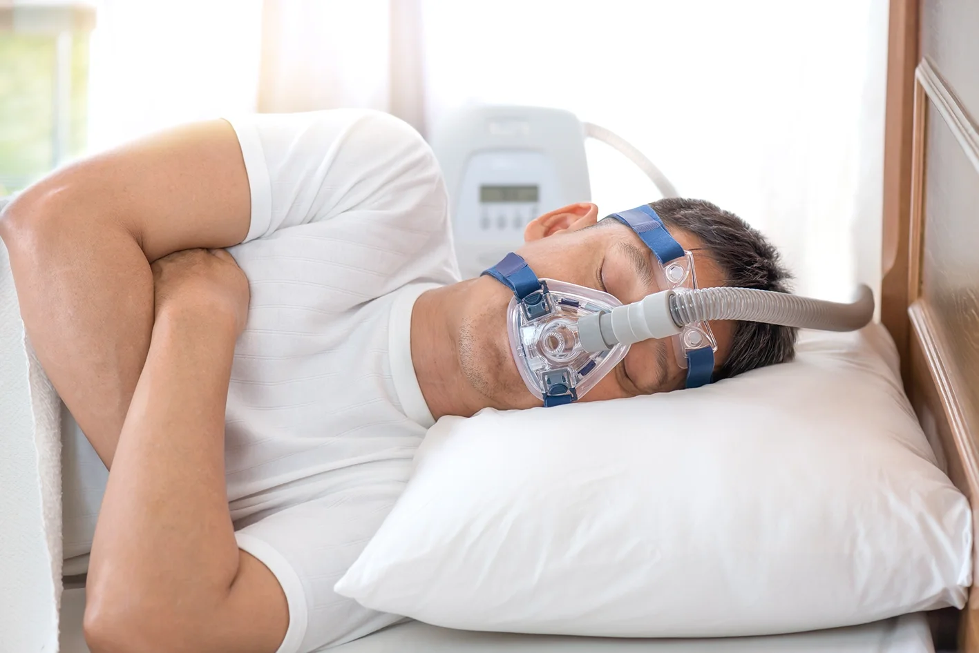
Continuous positive airway pressure, or CPAP, remains the standard treatment for many patients. However, maxillofacial surgery may provide an effective alternative for selected patients whose airway obstruction is related to jaw position or facial skeletal anatomy, particularly when CPAP or oral appliance therapy is not tolerated or does not provide adequate control.
Associate Professor Christian Freudlsperger provides specialist assessment and surgical treatment for obstructive sleep apnoea, with particular expertise in orthognathic surgery, maxillomandibular advancement and three-dimensional airway planning.
UNDERSTANDING THE CONDITION
What is Obstructive Sleep Apnoea (OSA)?
During sleep, the muscles supporting the tongue, soft palate and throat relax. In patients with OSA, the airway becomes partially or completely obstructed, causing:
Apnoeas, where breathing stops temporarily
Hypopnoeas, where airflow becomes significantly reduced
Repeated drops in blood oxygen
Brief awakenings that fragment normal sleep
These events may occur many times each hour without the patient being fully aware of them.
The severity of OSA is commonly measured using the apnoea–hypopnoea index, or AHI, obtained from a sleep study.
Health consequences of untreated OSA
Untreated moderate or severe OSA may contribute to:
High blood pressure
Heart disease
Heart attack
Stroke
Abnormal heart rhythms
Type 2 diabetes
Depression and reduced quality of life
Cognitive impairment
Increased risk of workplace and motor vehicle accidents
Effective treatment is therefore aimed not only at improving snoring and sleep quality, but also at reducing the wider health risks associated with repeated airway obstruction and oxygen desaturation.Anatomical factors contributing to OSA
Signs & Symptoms
Symptoms may include:
Loud or persistent snoring
Witnessed breathing pauses
Gasping or choking during sleep
Restless or unrefreshing sleep
Excessive daytime sleepiness
Morning headaches
Reduced concentration or memory
Irritability or mood changes
Dry mouth on waking
Frequent urination during the night
Reduced work or driving performance
A sleep study is important because symptoms alone cannot reliably determine the presence or severity of OSA. Polysomnography in a sleep laboratory remains the diagnostic gold standard for many clinical situations.
Anatomical Factors Contributing to OSA
Airway obstruction may be influenced by:
A small or recessed lower jaw
A deficient upper jaw or midface
Reduced space behind the tongue
A narrow upper jaw
Facial skeletal retrusion
Enlarged tongue or soft tissues
Enlarged tonsils
A long or thick soft palate
Nasal obstruction
Increased neck or upper-airway soft tissue
Obesity
More than one level of obstruction may be present. This is why surgical assessment must consider the nose, soft palate, tongue base, jaws and complete upper airway rather than focusing on a single structure.
PROCEDURE & TREATMENT OPTIONS
Comprehensive Sleep Assessment
Assessment is usually coordinated with a sleep physician and may include:
Review of sleep-study results
Evaluation of the AHI and oxygen levels
Review of daytime symptoms and snoring
Assessment of CPAP or oral appliance use
Facial and jaw examination
Dental bite and occlusal assessment
Examination of the tongue, palate and tonsils
Review of nasal symptoms
CBCT or CT imaging of the jaws and airway
Digital dental and facial scans
Drug-induced sleep endoscopy where appropriate
Discussion with an ENT surgeon, orthodontist or restorative dentist
Initial investigation commonly progresses from history and clinical examination to screening polygraphy and, where needed, formal polysomnography.
Non-Surgical Treatment Options
Surgery is not the first or only treatment for every patient. Non-surgical options may include:
CPAP therapy
A mandibular advancement splint
Weight management
Positional therapy
Treatment of nasal obstruction
Reduction of alcohol intake
Avoidance of sedating medication where medically appropriate
Improvement of sleep habits
CPAP remains the standard treatment for many patients with moderate or severe OSA. Surgery is considered when conservative therapy is unsuccessful, poorly tolerated or when a clear anatomical cause can be addressed.
SURGICAL TREATMENT OPTIONS
Maxillomandibular advancement
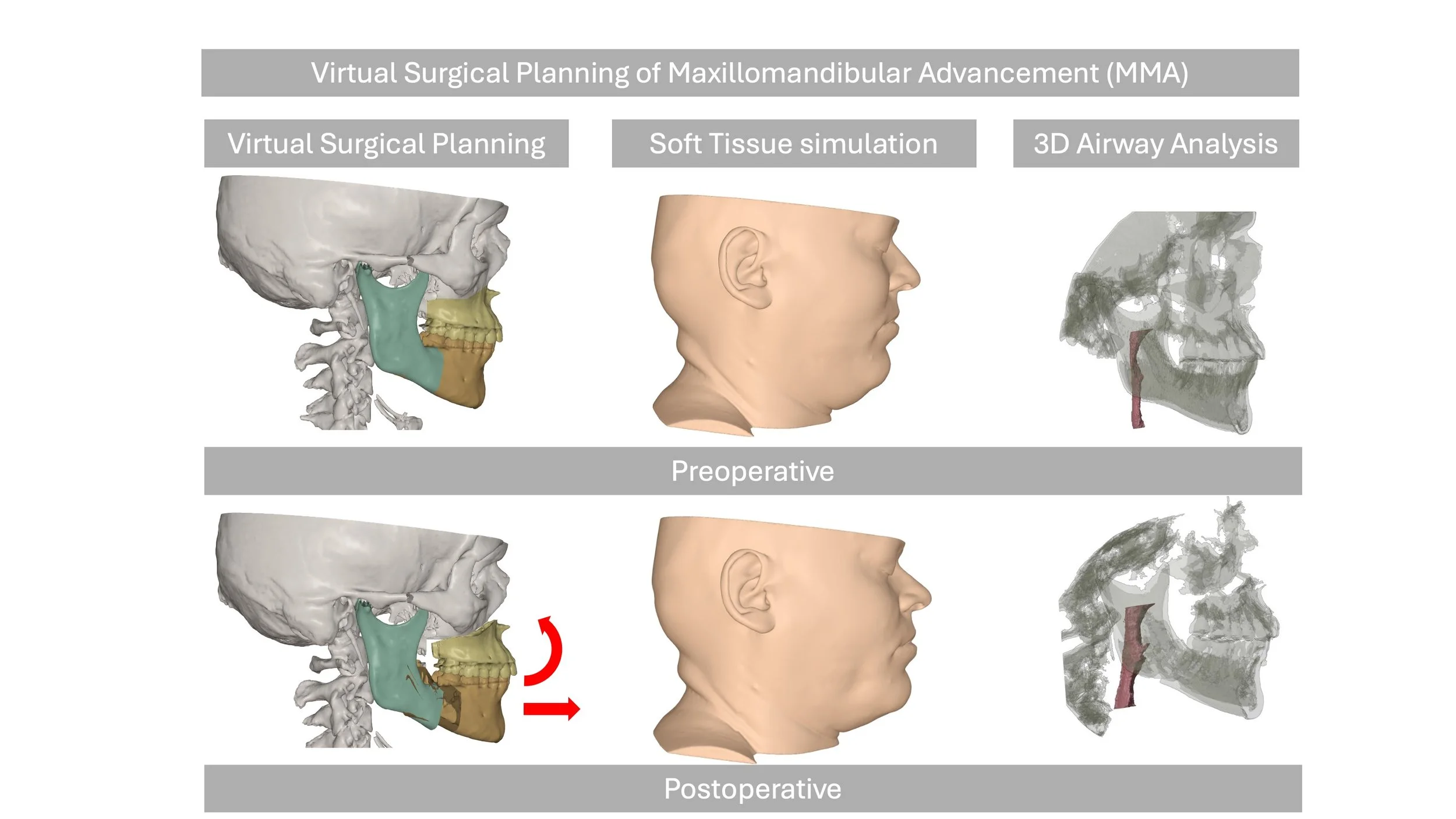
Maxillomandibular advancement, or MMA, moves both the upper and lower jaws forward. This increases the space behind the soft palate and tongue and improves tension within the surrounding airway tissues.
MMA is often considered for patients with:
Moderate or severe OSA
A recessed upper or lower jaw
Reduced posterior airway space
Failure or intolerance of CPAP
Inadequate response to an oral appliance
OSA associated with a significant bite or jaw discrepancy
Multilevel airway obstruction
It is one of the most effective surgical procedures for appropriately selected patients with skeletal airway restriction.
A/Prof Freudlsperger specializes in maxillomandibular advancement, which addresses the underlying skeletal deficiency causing airway compromise and provides the most predictable, long-lasting results.
The Surgical Process
Maxillomandibular advancement is performed under general anaesthesia in hospital.
The procedure generally involves:
Repositioning the upper jaw with a Le Fort I osteotomy
Repositioning the lower jaw with a bilateral sagittal split osteotomy
Advancing both jaws according to the digital plan
Stabilising the jaws with titanium plates and screws
Using guiding elastics to support the bite during healing
The incisions are made inside the mouth, so visible facial scars are generally avoided.
Most patients remain in hospital for two nights. Traditional prolonged jaw wiring is not routinely required in most modern cases.
Why Choose A/Prof Christian Freudlsperger?
Specialist Orthognathic and Airway Expertise: Associate Professor Christian Freudlsperger has extensive experience in corrective jaw surgery and maxillomandibular advancement, allowing airway treatment to be planned together with bite function and facial balance.
Advanced 3D Planning: Digital jaw, dental and airway imaging support precise simulation of skeletal movements and coordinated surgical planning.
Comprehensive Anatomical Assessment: Treatment considers the entire upper airway, including the nose, palate, tongue base, jaws and facial skeleton.
Multidisciplinary Collaboration: Care is coordinated with sleep physicians, respiratory specialists, ENT surgeons, orthodontists and dentists to ensure that surgery is appropriate and forms part of a complete OSA treatment plan.
RISK & RECOVERY
Understanding the Risks
Surgical treatment of OSA carries risks similar to orthognathic surgery: swelling (significant but temporary), numbness or altered sensation (common initially, usually improves over months), infection (uncommon), bleeding, jaw joint discomfort, bite changes (usually managed with short-term orthodontics), and general anaesthesia risks. Treatment failure or incomplete resolution of OSA can occur but is less common with MMA surgery than other surgical approaches.
A/Prof Freudlsperger will discuss all potential risks, benefits, and alternatives specific to your individual case in detail during your consultation, ensuring you have a complete understanding before proceeding with treatment.
What to Expect During Recovery
Hospital Stay: 2 nights for monitoring.
First Two Weeks: Significant swelling, liquid/soft diet, discomfort managed with medication, limited activity, and sleep with head elevated. Despite swelling, many patients notice immediate improvement in breathing.
Weeks 3-6: Swelling decreasing, gradual return to normal diet and activities, continued improvement in symptoms, and follow-up sleep study typically scheduled around 3-6 months post-surgery.
Months 3-12: Continued subtle improvements in swelling and sensation, achievement of final results, and documented improvement in sleep study parameters.
Post-Operative Care
Following post-operative instructions is essential: maintain excellent oral hygiene, follow dietary progression, take prescribed medications, avoid strenuous activity initially, no smoking (severely impairs healing and worsens OSA), sleep with head elevated initially, attend all follow-up appointments, and complete follow-up sleep study to document improvement.
Most patients experience dramatic improvement in sleep quality, daytime energy, and overall health following successful surgical treatment.
Frequently Asked Questions
1. Can surgery cure obstructive sleep apnoea?
Surgery can produce a major improvement and may normalise sleep-study results in some carefully selected patients. However, no procedure can guarantee a cure.
The outcome depends on OSA severity, anatomy, body weight, the level of airway obstruction and the operation performed. A postoperative sleep study is required to confirm the result.
2. What is the most effective surgery for sleep apnoea?
For patients whose OSA is related to recessed jaws or reduced skeletal airway support, maxillomandibular advancement is among the most effective surgical options.
Other procedures may be more appropriate when obstruction is primarily related to the tonsils, nose, palate or another specific structure.
3. Do I need to try CPAP before considering surgery?
Usually, CPAP is recommended first, particularly for moderate or severe OSA. Surgery may be considered when CPAP is not tolerated, does not provide adequate control or when there is a clear skeletal abnormality that can be corrected.
The decision is made with the sleep physician and surgical team.
4. Will jaw advancement change my face?
Yes. Moving the jaws forward changes the facial profile, lips and chin position.
These changes are carefully planned. For patients with recessed jaws, advancement often improves facial balance as well as airway support. Three-dimensional planning helps assess the functional and facial effects before treatment.
5. Will I still need CPAP after sleep apnoea surgery?
Some patients no longer require CPAP after successful surgery, while others may still need CPAP at a lower pressure or use an oral appliance.
Treatment should not be stopped until a follow-up sleep study has confirmed that the OSA has improved sufficiently.

BOOK YOUR APPOINTMENT
Breathe Better, Sleep Better
If you have moderate or severe obstructive sleep apnoea, difficulty tolerating CPAP, a recessed jaw, reduced airway space or persistent symptoms despite previous treatment, please contact Oral & Facial Surgery to arrange a specialist assessment with Associate Professor Christian Freudlsperger.
We welcome referrals from sleep physicians, respiratory specialists, ENT surgeons, orthodontists, dentists and general practitioners.