

TMJ Surgery
Specialist treatment for jaw joint pain, locking and advanced TMJ disease
Temporomandibular disorders, commonly called TMD or TMJ disorders, are a group of conditions affecting the temporomandibular joints, chewing muscles and associated tissues. Symptoms may include persistent jaw pain, clicking, locking, headaches, restricted mouth opening and difficulty eating or speaking.
TMD is common, but only a small proportion of patients require surgery. Many symptoms improve with appropriate conservative treatment, and some resolve spontaneously. The attached presentation notes that only approximately 5–10% of symptomatic patients require surgical management, reinforcing the importance of careful diagnosis and appropriate patient selection.
Associate Professor Christian Freudlsperger has performed TMJ surgery since 2011 and provides the complete spectrum of surgical treatment, including arthrocentesis, TMJ arthroscopy, open joint surgery and total temporomandibular joint replacement.
He has more than 20 years of international experience in oral and maxillofacial surgery, with a particular clinical and academic focus on advanced TMJ reconstruction and total joint replacement. His work has included research, international teaching and collaboration with industry partners in the development and refinement of TMJ prosthetic concepts.
TMJ surgery should always be considered carefully. The expected benefits must be weighed against the risks, limitations and non-surgical alternatives. Advanced procedures should be undertaken by surgeons experienced in jaw-joint surgery, facial anatomy and complex reconstruction.
UNDERSTANDING THE CONDITION
What are the Temporomandibular Joints?
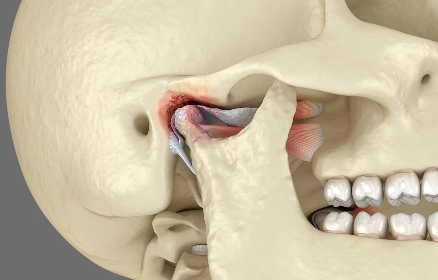
The temporomandibular joints connect the lower jaw to the skull immediately in front of each ear. They are complex joints that combine rotational and sliding movements, allowing the jaw to open, close and move from side to side.
Each joint contains a cartilage disc positioned between the mandibular condyle and the temporal bone. The disc helps distribute pressure and supports smooth movement.
TMD may involve:
The joint surfaces and cartilage disc
The joint capsule and synovial lining
The chewing muscles
Supporting ligaments
The dental bite and jaw relationship
The surrounding pain-processing system
Types of TMJ Disorders
Myogenic TMD
Myogenic TMD primarily affects the chewing muscles rather than the joint itself.
It may be associated with:
Clenching or grinding
Muscle overuse
Stress and tension
Altered jaw posture
Neck and shoulder dysfunction
Chronic pain sensitisation
Myogenic TMD is usually treated conservatively. Joint surgery does not generally help pain that originates mainly from the muscles.
Intra-Articular Pain and Dysfunction
Intra-articular pain and dysfunction arises from within the joint.
Current understanding recognises that excessive mechanical loading may trigger inflammatory changes within the synovial membrane. This can produce pain, swelling, restricted movement and progressive joint degeneration.
Possible causes include:
Synovitis
Capsular impingement
Fibrous adhesions
Symptomatic disc displacement
Cartilage damage
Osteoarthritis
Disc displacement on an MRI does not automatically mean that the disc is causing the patient’s symptoms. A mechanical imaging finding must be interpreted alongside the clinical examination.
Internal Derangement
Internal derangement refers to abnormal movement or position of the joint disc.
Symptoms may include:
Painful clicking or popping
Intermittent locking
Sudden reduction in mouth opening
Deviation of the jaw
Pain during chewing or yawning
A displaced disc may click as it moves back into position. If it remains displaced, the joint may become painful or locked.
Degenerative Joint Disease
Osteoarthritis and inflammatory arthritis may damage the cartilage and bone of the TMJ.
Symptoms can include:
Persistent joint pain
Grating or crunching sounds
Progressive loss of movement
Changes in the bite
Loss of joint height
Facial asymmetry
Ankylosis
TMJ ankylosis occurs when the joint becomes partly or completely fused by fibrous tissue or bone.
It may follow:
Trauma
Infection
Severe arthritis
Previous surgery
Childhood condylar injury
Ankylosis can cause profound restriction of mouth opening and may affect facial growth in younger patients.
End-Stage Joint Disease
Advanced joint destruction may occur following severe arthritis, ankylosis, trauma or multiple previous operations.
In appropriately selected patients, total TMJ replacement may provide the most predictable method of restoring movement, reducing pain and rebuilding the joint.
Signs & Symptoms
Assessment may be appropriate for:
Persistent pain over the jaw joint
Painful clicking, popping or grinding
Recurrent jaw locking
Reduced or progressively worsening mouth opening
Difficulty chewing or speaking
Deviation of the jaw during opening
Changes in the bite
Facial asymmetry
Pain following previous surgery or trauma
Severe degenerative changes on imaging
TMD may also produce pain around the ear, temple, eye, neck, shoulder or side of the face. However, facial pain perceived as “TMJ pain” may originate from another source, making accurate diagnosis essential.
PROCEDURE & TREATMENT OPTIONS
Comprehensive TMJ Assessment
Diagnosis is the most important step in successful TMJ treatment.
The assessment aims to distinguish:
Muscular or myogenic pain
Joint-related or arthrogenic pain
Dental pain
Ear-related symptoms
Neuropathic facial pain
Headache disorders
Inflammatory arthritis
Structural joint disease
Examination may include:
A detailed history of pain, clicking and locking
Measurement of pain-free and assisted mouth opening
Assessment of lateral and forward jaw movement
Observation of jaw deviation
Palpation of the TMJs
Examination of the chewing and neck muscles
Assessment of the ear canal and eardrum where relevant
Evaluation of the bite
Assessment of joint sounds
Review of previous splints, physiotherapy, injections or surgery
The presentation emphasises an “outside-to-inside” examination, including assessment of possible ear-related causes, muscle palpation, joint palpation, movement measurement and occlusal examination.
Imaging
Imaging is selected according to the suspected diagnosis.
Panoramic Radiography
An OPG or panoramic X-ray may be used as an initial investigation to assess the teeth, jaws and obvious bony joint abnormalities.
Dental conditions are an important potential source of facial pain and must be excluded.
CT or CBCT
CT or CBCT provides detailed imaging of:
The condyle and joint socket
Osteoarthritis
Bony ankylosis
Fracture or deformity
Previous surgical changes
Bone loss or overgrowth
Planning for total joint replacement
MRI
MRI is the preferred investigation for:
Disc position and shape
Joint fluid or effusion
Synovial inflammation
Soft-tissue abnormalities
Internal derangement
Closed- and open-mouth MRI images can demonstrate how the disc and condyle move during jaw opening.
Imaging findings are interpreted alongside the patient’s symptoms and examination. An abnormal scan alone is not an indication for surgery.
NON-SURGICAL TREATMENT
Conservative Treatment Approaches
Most patients should receive an appropriate period of non-surgical treatment before surgery is considered.
Treatment may include:
Education and supported self-care
Temporary soft diet
Heat or cold therapy
Avoidance of gum chewing and excessive opening
Identifying and reducing clenching or grinding
Jaw exercises
Physiotherapy
Anti-inflammatory or pain medication
A stabilisation splint where appropriate
Stress-management strategies
Cognitive behavioural approaches
Management of inflammatory arthritis
Pain-specialist or oral-medicine input
The presentation recommends a defined period of conservative treatment, commonly around six to eight weeks, for joint-related symptoms before escalation, while avoiding prolonged ineffective treatment when a clearly defined intra-articular disorder persists.
SURGICAL PROCEDURES
Surgical Treatment Options
When conservative treatments fail to provide adequate relief, surgical options include:
Arthrocentesis and Joint Lavage
Arthrocentesis is a minimally invasive procedure commonly used as an initial surgical treatment for a painful or locked TMJ.
Two small needles are introduced into the upper joint space, and sterile fluid is circulated through the joint.
This may:
Remove inflammatory mediators and degraded proteins
Reduce joint pressure
Improve mobility
Release minor adhesions
Reduce pain and stiffness
Improve mouth opening
The presentation reports published improvement in pain and range of movement in approximately 70–95% of appropriately selected patients, although individual outcomes vary.
Arthrocentesis may be performed under local anaesthetic, intravenous sedation or general anaesthesia.
Physiotherapy and postoperative jaw exercises are important to maintain movement.
TMJ Arthroscopy
TMJ arthroscopy is a minimally invasive keyhole procedure in which a small camera and instruments are introduced into the upper joint space.
It allows the surgeon to inspect the joint directly and treat abnormalities such as:
Synovitis
Capsular impingement
Fibrous adhesions
Synovial folds
Chondromalacia
Restricted disc movement
Arthroscopy may be used to:
Lavage the joint
Release adhesions
Remove inflamed tissue
Improve joint movement
Reduce pain and locking
The presentation reports published improvement in pain and movement in approximately 80–90% of selected patients. It also notes that arthroscopy may provide better outcomes than arthrocentesis in some groups because physical abnormalities can be treated directly.
Arthroscopy requires specialised equipment and surgical training and has a significant learning curve.
Open Joint Surgery - Arthroplasty
Open TMJ surgery may be considered when a defined structural abnormality cannot be treated adequately with conservative or minimally invasive procedures.
Possible indications include:
Severely damaged or displaced disc
Persistent mechanical locking
Advanced degenerative disease
Bony overgrowth
Loose bodies
Ankylosis
Condylar deformity
Benign joint lesions
Failure of previous minimally invasive treatment
Procedures may include:
Disc repositioning or repair
Discectomy
Removal of adhesions or scar tissue
Eminoplasty
Condylar reshaping
Condylectomy
Ankylosis release
Joint reconstruction
If minimally invasive treatment fails to improve pain and function, open arthroplasty or total joint replacement may be considered according to the severity and reversibility of the disease.
Repeated unsuccessful open procedures may make future treatment more difficult and may reduce the predictability of later reconstruction. For this reason, each operation should have a clearly defined diagnosis and objective.
Total TMJ Replacement
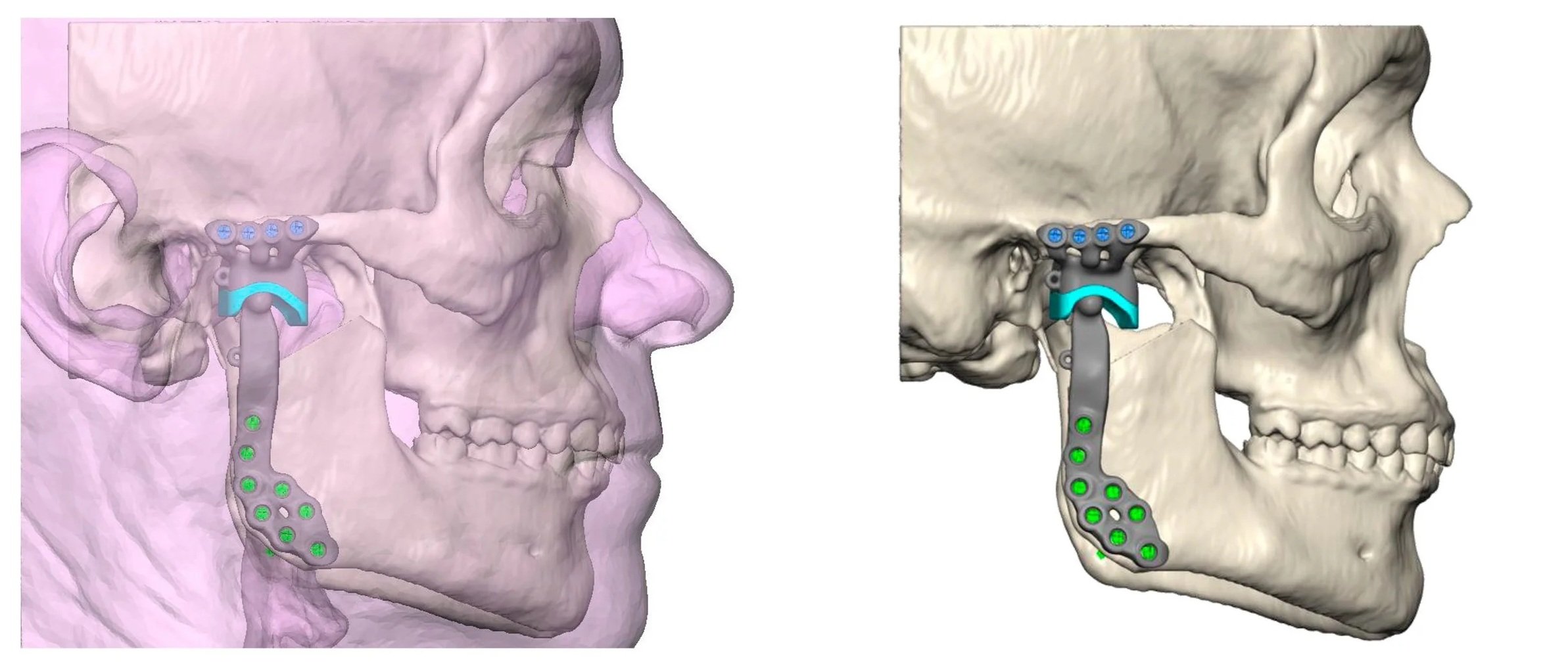
Total TMJ replacement replaces the damaged mandibular condyle and joint socket with prosthetic components.
It is reserved for severe, irreversible joint disease and is not an initial treatment for routine jaw pain, clicking or uncomplicated disc displacement.
TOTAL TMJ REPLCEMENT SURGERY
When Is Total TMJ Replacement Considered?
Possible indications include:
End-stage osteoarthritis
Severe inflammatory arthritis
Advanced joint destruction
Bony or recurrent ankylosis
Failed previous TMJ surgery
Severe condylar deformity or loss
Major functional restriction
Persistent joint pain associated with irreversible structural disease
Reconstruction following removal of diseased joint tissue
Planning Total TMJ Replacement
Assessment may include:
High-resolution CT imaging
Digital three-dimensional reconstruction
Evaluation of the bite and facial symmetry
Review of previous surgery and implants
Assessment of the facial nerve
Dental and medical optimisation
Virtual surgical planning
Planning for simultaneous orthognathic surgery where required
Custom-made joint components may be designed from the patient’s CT scan to match the temporal bone and lower jaw.
Associate Professor Freudlsperger has a particular focus on total TMJ replacement, including complex ankylosis, failed previous surgery and joint-related facial deformity. He has been involved in research, international lectures and collaboration with industry partners concerning the design and development of TMJ prostheses.
What can Total TMJ Replacement achieve?
Potential benefits include:
Improved mouth opening
Reduced joint pain
Improved chewing
Restoration of mandibular continuity
Correction of joint-related deformity
Improved bite stability
Reduced risk of recurrent bony ankylosis
Improved quality of life
A prosthetic joint cannot reproduce all features of a natural TMJ. Expectations, limitations and the possibility of future revision must be discussed carefully.
A/Prof Freudlsperger carefully evaluates each patient to determine the most appropriate treatment approach, emphasizing conservative management first and recommending surgery only when clearly indicated.
Why Choose A/Prof Christian Freudlsperger?
Comprehensive TMJ Surgical Expertise: Associate Professor Christian Freudlsperger has performed TMJ surgery since 2011 and provides the full surgical spectrum from arthrocentesis and arthroscopy to open reconstruction and total joint replacement.
Specialist Focus on Total Joint Replacement: Advanced joint destruction, ankylosis and failed previous surgery require expertise in TMJ reconstruction, facial skeletal surgery and three-dimensional planning.
Diagnosis-Driven Treatment: Particular emphasis is placed on distinguishing myogenic pain from true intra-articular disease before considering surgery.
Careful Surgical Decision-Making: Surgery is recommended only when the diagnosis, imaging, functional limitation and response to previous treatment indicate that the expected benefits outweigh the risks.
Multidisciplinary Collaboration: Care may be coordinated with dentists, orthodontists, physiotherapists, rheumatologists, pain specialists and medical practitioners.
RISKS & RECOVERY
Understanding the Risks
Conservative treatment risks are minimal (possible medication side effects, temporary soreness from exercises). Surgical treatment carries risks depending on the procedure: infection (uncommon, treated with antibiotics), increased pain or limited improvement (occasional non-response), nerve injury causing numbness or weakness (rare), hearing changes (joint proximity to ear), need for additional procedures, and general anesthesia risks for open procedures.
A/Prof Freudlsperger will discuss all potential risks, benefits, and alternatives specific to your individual case in detail during your consultation, ensuring you have a complete understanding before proceeding with treatment.
What to expect during your recovery
Arthrocentesis:
Mild swelling and tenderness may continue for several days. Most patients return to light activities quickly.
Jaw exercises usually begin shortly after treatment.
TMJ Arthroscopy:
A soft diet and regular exercises are recommended initially. Swelling and discomfort generally improve over several days.
Open Joint Surgery:
Recovery commonly includes swelling and tenderness for one to two weeks, a soft diet and structured physiotherapy.
Return to usual activities generally occurs progressively over four to six weeks.
Total Joint Replacement:
Joint replacement requires hospital care and a longer rehabilitation programme.
Recovery may include:
Early postoperative imaging
Liquid and soft-food progression
Regular mouth-opening exercises
Physiotherapy
Monitoring of the bite
Facial nerve review
Long-term clinical and radiographic surveillance
Improvement in function occurs gradually, while swelling and altered sensation may continue to settle over several months.
Post-Operative Care
For surgical procedures, success depends on following instructions: take prescribed medications, follow dietary restrictions, perform prescribed jaw exercises consistently, apply ice or heat as directed, maintain excellent oral hygiene, avoid habits that stress the jaw (gum chewing, nail biting), attend regular physical therapy and follow-up appointments, and manage stress and address grinding/clenching habits.
Many patients experience significant improvement in pain and function with appropriate treatment, though some chronic conditions require ongoing management.
Frequently Asked Questions
1. Do all TMJ disorders need surgery?
No. Most TMD is treated conservatively, and only a minority of patients require surgery.
Surgery is considered when a defined joint disorder causes persistent pain, locking or functional limitation despite appropriate treatment.
2. What is the difference between muscular TMD and joint-related TMD?
Muscular TMD causes pain mainly in the chewing muscles and is often associated with clenching, grinding or tension.
Joint-related TMD causes pain or dysfunction within the TMJ itself. The distinction is important because joint surgery does not generally improve primarily muscular pain.
3. What is the difference between arthrocentesis and arthroscopy?
Arthrocentesis washes the joint through small needles.
Arthroscopy uses a small camera and instruments, allowing the surgeon to inspect the joint and treat adhesions, inflamed tissue and other internal abnormalities directly.
4. Can TMJ surgery stop clicking and pain?
Surgery may reduce pain, locking and mechanical symptoms when these arise from a clearly diagnosed joint disorder.
Painless clicking alone rarely requires surgery. Muscle pain, clenching and chronic pain sensitisation may not improve with joint surgery.
5. When is total TMJ replacement necessary?
Total joint replacement is considered for severe irreversible joint destruction, ankylosis, major deformity or failed previous surgery.
It is not usually appropriate for early disc displacement, uncomplicated clicking or muscular TMD.
BOOK YOUR APPOINTMENT
Find Relief from TMJ Pain
Referrals are welcome for:
Persistent or recurrent TMJ locking
Joint-related pain that has not responded to appropriate treatment
Disc displacement and internal derangement
Degenerative or inflammatory joint disease
Ankylosis
Condylar deformity or loss
Previous unsuccessful TMJ surgery
Assessment for open joint reconstruction
Assessment for total TMJ replacement
Referrals are welcomed from dentists, orthodontists, oral medicine specialists, physiotherapists, pain specialists, rheumatologists, GPs and other medical practitioners.