

Reconstructive Surgery
Advanced jaw reconstruction, virtual surgical planning and dental rehabilitation
Reconstructive oral and maxillofacial surgery restores the structure, function and appearance of the jaws and face after cancer treatment, benign jaw tumours, osteonecrosis, infection, trauma or previous unsuccessful surgery.
Reconstruction is planned around the patient’s long-term needs from the outset. The aim is not simply to replace missing bone or soft tissue, but to restore jaw continuity, facial contour, speech, swallowing, chewing and, where possible, fixed dental function with implant-supported teeth.
Associate Professor Christian Freudlsperger provides specialist assessment and treatment for complex reconstruction of the mandible, maxilla and midface. He has more than 20 years of international experience in oral and maxillofacial, oncological and microvascular reconstructive surgery, with particular expertise in virtual surgical planning, patient-specific reconstruction and dental rehabilitation after jaw reconstruction.
His ongoing academic work includes virtual planning of mandibular reconstruction, and he is a co-author of recent research into automated knowledge-based VSP for mandibular defects. He also chairs international advanced surgical training in free-fibula mandibular reconstruction with immediate dental implant planning.
UNDERSTANDING THE PROCEDURE
When is reconstructive surgery needed?
Reconstruction of the lower jaw, upper jaw or facial skeleton may be required following:
Oral or head and neck cancer surgery
Benign but locally aggressive jaw tumours, including ameloblastoma
Large jaw cysts or fibro-osseous lesions
Osteoradionecrosis following radiotherapy
Medication-related osteonecrosis of the jaw
Chronic osteomyelitis or severe infection
Facial trauma with loss of bone or soft tissue
Congenital or developmental conditions
Failed plates, grafts, implants or previous reconstruction
Significant deformity following earlier surgery
Jawbone loss preventing dental rehabilitation
These are recognised indications for microvascular bone reconstruction of the mandible and maxilla.
What Are the Goals of Jaw Reconstruction?
The objectives depend on the defect and the patient’s priorities but may include restoring:
Jaw continuity and skeletal stability
Facial contour and symmetry
A stable relationship between the upper and lower jaws
Speech and tongue mobility
Safe swallowing
Separation of the mouth from the nose or sinuses
Lip competence and oral closure
Chewing function
Bone suitable for dental implants
Appearance, confidence and quality of life
Loss of mandibular continuity can affect the bite, chewing, swallowing, airway and lower facial contour. Successful reconstruction therefore needs to reproduce the three-dimensional shape of the jaw and provide stable support for the surrounding soft tissues.
YOUR PROCEDURE
Comprehensive Assessment and Treatment Planning
Reconstructive treatment begins with a detailed assessment of the underlying disease, existing defect, general health and long-term functional goals.
Planning may include:
Clinical examination of the face, mouth and neck
Assessment of speech, swallowing and tongue mobility
Evaluation of the teeth, bite and jaw joints
CT or CBCT imaging of the facial skeleton
CT angiography of a potential donor site
Digital intraoral scans
Facial photography or three-dimensional facial scanning
Review of previous surgery, radiotherapy and pathology
Prosthodontic assessment for future teeth
Multidisciplinary cancer review where appropriate
Where reconstruction follows cancer surgery, the planned tumour resection, reconstructive procedure and need for postoperative radiotherapy must be considered together.
Virtual Surgical Planning
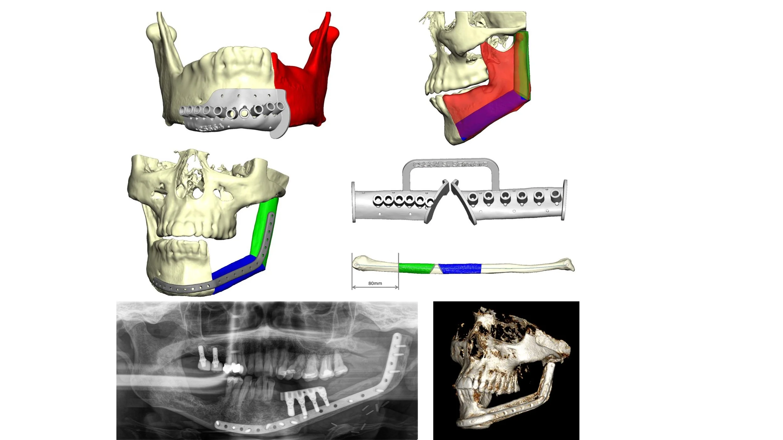
Virtual surgical planning, or VSP, creates a three-dimensional digital model of the patient’s jaw, defect and proposed reconstruction before surgery.
The process may involve:
Digitally defining the area of bone that requires removal
Reconstructing the expected jaw shape in three dimensions
Selecting and positioning the most appropriate donor bone
Planning the bone cuts required to shape the graft
Positioning the reconstructed upper and lower jaws in the correct relationship
Planning future dental implant positions
Producing customised cutting guides, models and fixation components
The digital plan can be transferred to surgery using three-dimensionally printed resection guides, donor-bone cutting guides and patient-specific titanium implants.
Associate Professor Freudlsperger’s published work describes how VSP and CAD/CAM technology can improve reconstructive precision, predictability, bone positioning and the relationship between the reconstructed jaws.
Patient-Specific Reconstruction
In selected complex cases, a customised titanium reconstruction plate or patient-specific implant can be manufactured from the digital plan.
Potential advantages include:
More accurate restoration of the original jaw shape
Precise positioning of transferred bone segments
Improved alignment of the upper and lower jaws
Better preparation for later dental implants
Reduced intraoperative adjustment
Improved facial contour and symmetry
Greater predictability in secondary or revision reconstruction
Published evidence reviewed by Associate Professor Freudlsperger and colleagues describes increased accuracy and close agreement between the virtual plan and postoperative reconstruction.
REHABILITATION & RECOVERY
Dental implant rehabilitation after jaw reconstruction
Dental rehabilitation is considered an integral part of jaw reconstruction rather than an optional final step.
Where suitable, implants may be placed:
At the time of jaw reconstruction
During a planned second procedure after healing
After radiotherapy and soft-tissue stabilisation
As part of secondary reconstruction of an existing defect
The timing depends on:
The diagnosis and risk of recurrence
Need for radiotherapy
Quality and position of reconstructed bone
Soft-tissue condition
Implant stability
Medical fitness
The planned prosthesis
Rebuilding adequate bone and preserving the correct relationship between the upper and lower jaws are essential for implant-supported dental rehabilitation.
Immediate implant rehabilitation and “Jaw in a Day”
In highly selected cases, dental implants can be incorporated into the virtual reconstruction plan and placed into the bone flap during the jaw reconstruction procedure.
This may shorten the interval between reconstruction and fixed dental rehabilitation. However, immediate implant placement is not appropriate for every patient and requires close coordination between the reconstructive surgeon, prosthodontist and dental laboratory.
Associate Professor Freudlsperger has published on fully three-dimensional mandibular reconstruction using a vascularised fibula and implant-supported prosthetic rehabilitation
Recovery
Recovery timeframes vary considerably depending on the extent and type of reconstruction. Smaller grafting procedures may involve a recovery period of several weeks, while more extensive reconstruction — particularly involving tissue transfer — requires a longer period of healing and structured follow-up, often over several months, to monitor integration and function.
Why Choose Oral & Facial Surgery Remeura?
Specialist Jaw Reconstruction Expertise: Associate Professor Christian Freudlsperger has extensive experience in reconstruction of the mandible, maxilla and midface following cancer, benign tumours, osteonecrosis, infection, trauma and previous unsuccessful surgery.
International Experience in Virtual Surgical Planning: His clinical and academic work includes VSP, fibula and iliac-crest reconstruction, patient-specific implants and implant-supported rehabilitation. His recent research affiliation spans Heidelberg University Hospital and the Oral and Maxillofacial Surgery Service at Te Toka Tumai Auckland.
Reconstruction Designed Around Dental Rehabilitation: The intended bite, implant positions and final prosthesis are considered during surgical planning so that the reconstructed jaw supports future chewing and fixed tooth replacement wherever possible.
Comprehensive Multidisciplinary Care: Complex treatment is coordinated with head and neck surgeons, oncologists, radiologists, pathologists, prosthodontists, restorative dentists, speech-language therapists, dietitians and specialist nurses.
Frequently Asked Questions
1. Is jaw reconstruction mainly cosmetic or functional?
It is both, but restoration of function is the primary objective. Reconstruction aims to restore jaw stability, chewing, speech, swallowing and oral closure while also rebuilding facial contour and symmetry.
2. What is virtual surgical planning for jaw reconstruction?
Virtual surgical planning uses CT scans and digital models to simulate the bone removal and reconstruction before surgery. Custom cutting guides and patient-specific fixation can then transfer the plan accurately to the operating theatre.
3. Which bone is used to reconstruct the jaw?
The fibula, iliac crest and scapula are the principal donor sites for vascularised bone reconstruction. The choice depends on the size and shape of the defect, required soft tissue, dental rehabilitation goals and the patient’s general health.
4. Can dental implants be placed into a reconstructed jaw?
Yes. Dental implants can often be placed into reconstructed fibula, iliac-crest or scapular bone. Suitability depends on bone position and volume, soft-tissue quality, radiotherapy, medical health and the intended prosthesis.
5. Can implants be placed at the same time as jaw reconstruction?
In selected patients, implants may be placed during the reconstruction. This is sometimes described as immediate dental rehabilitation or a “jaw-in-a-day” approach. Careful patient selection and coordinated digital planning are essential.
6. How long does jaw reconstruction take to recover from?
Initial recovery from major reconstruction generally takes several weeks, but complete healing and rehabilitation take longer. Dental restoration may require staged treatment over several months, particularly following cancer surgery or radiotherapy.
7. Can a jaw be reconstructed after radiotherapy or osteonecrosis?
Yes, but irradiated or infected tissue requires specialist assessment. Advanced osteoradionecrosis or medication-related osteonecrosis may require removal of diseased bone and reconstruction with healthy vascularised tissue.
A collaborative approach
Complex reconstructive cases are often managed with input from other specialists, including plastic surgeons, ENT surgeons, oncologists and prosthodontists, to achieve the best possible functional and aesthetic outcome for each patient.
Next steps
If you require reconstruction of the mandible, maxilla or midface following cancer, ameloblastoma, osteoradionecrosis, medication-related osteonecrosis, chronic infection, trauma or previous failed surgery, please contact Oral & Facial Surgery to arrange a specialist consultation with Associate Professor Christian Freudlsperger.
Please bring any available scans, pathology results, operation reports and specialist correspondence to your appointment.
Referrals are welcome from oral and maxillofacial surgeons, head and neck surgeons, dentists, prosthodontists, orthodontists, oncologists, radiation oncologists, GPs and other medical specialists.
BOOK YOUR APPOINTMENT
Schedule Your Consultation
Contact our rooms to arrange a comprehensive consultation with A/Prof Freudlsperger. During this consultation, he will provide honest assessment of what can be achieved with your unique facial anatomy.