

Oral Pathology
Early diagnosis of mouth changes
Persistent ulcers, lumps, swellings, red or white patches, unexplained bleeding, numbness or other changes in the lining of the mouth should not be ignored. Many oral conditions are benign, but some require prompt investigation and treatment.
Associate Professor Christian Freudlsperger and Mr Nigel Parr provide specialist assessment and surgical management of conditions affecting the mouth, jaws and salivary glands. Care ranges from clinical examination and minor biopsy procedures to treatment of oral potentially malignant disorders, benign tumours and oral cancer.
Early assessment is particularly important because oral cancer is frequently diagnosed at an advanced stage, while outcomes are substantially better when disease is identified early
UNDERSTANDING THE CONDITION
Types of Oral Pathology
Oral pathology includes a wide range of conditions affecting the lips, tongue, cheeks, gums, palate, floor of the mouth, jawbones and salivary glands.
Oral mucosal conditions
These include:
Persistent mouth ulcers
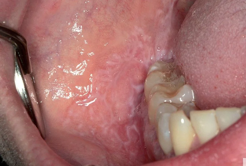
White patches, including leukoplakia
Red patches, including erythroplakia
Mixed red and white areas
Lumps, nodules or thickened tissue
Pigmented lesions
Oral lichen planus and lichenoid conditions
Inflammatory, autoimmune and infectious disorders
Some visible oral changes are classified as oral potentially malignant disorders. This means they have a variable risk of developing into oral cancer, although most do not become malignant. Cancer may occasionally develop elsewhere in the oral cavity as well as within the original lesion, which is why ongoing review can be important.
PROCEDURE & TREATMENT OPTIONS
Warning signs requiring evaluation
Please seek assessment for:
An ulcer that has not healed within two weeks
A persistent red, white or mixed red-and-white patch
A new lump, swelling or area of thickening
A lesion that is firm, fixed or changing in appearance
Unexplained oral pain, bleeding or numbness
Difficulty or pain when swallowing
A non-healing extraction socket
An unexplained loose tooth
A persistent neck lump
Any oral lesion causing significant concern
Persistent ulcerated, verrucous, red, white or mixed lesions, particularly those with induration, fixation, increasing pain or altered sensation, may require biopsy.
Risk factors for oral cancer
Important risk factors include:
Tobacco smoking or chewing tobacco
Heavy alcohol consumption, particularly when combined with tobacco
Previous oral cancer or potentially malignant disease
Human papillomavirus in cancers arising in the tonsils or base of tongue
Excessive sun exposure for lip cancer
Increasing age
Immunosuppression
Oral cancer can also occur in people without recognised risk factors. Regular dental examinations and review of persistent changes remain important.
ASSESSMENT & BIOPSY
Comprehensive Evaluation
Assessment begins with a detailed history and careful examination of the entire mouth. The neck may also be examined for enlarged lymph nodes or salivary gland abnormalities.
Depending on the findings, further assessment may include:
Clinical photographs to document and monitor the lesion
Dental radiographs or an OPG
Ultrasound for salivary gland or neck lesions
CBCT, CT or MRI for deeper or more complex conditions
Blood tests or microbiological investigations
Biopsy and histopathological examination
Clinical photographs and specialist referral are particularly helpful when a lesion changes or a new lesion develops.
Biopsy Procedures
A biopsy involves removing a small tissue sample so that it can be examined under a microscope by a specialist pathologist. Most biopsies are performed to clarify a diagnosis and do not mean that cancer is suspected.
Incisional biopsy: A representative sample is removed from a larger or more suspicious lesion. This provides a diagnosis before definitive treatment is planned.
Excisional biopsy: A smaller lesion is removed completely. This may provide both diagnosis and treatment if the condition is benign.
Punch biopsy: A small circular instrument is used to obtain a precise sample from the lining of the mouth.
Fine-needle aspiration: A fine needle may be used to collect cells or fluid from a salivary gland lesion, cyst or neck lump.
Most minor oral biopsies are performed under local anaesthetic. Sedation or general anaesthesia may be considered for larger, deeper or more complex lesions. Tissue is sent for histopathological analysis, and the result and recommended next steps are discussed once available.
PROCEDURE & TREATMENT OPTIONS
Treatment Approaches
Treatment is based on the confirmed diagnosis and may include:
Reassurance and observation
Removal of an irritating dental or local cause
Topical or systemic medication
Treatment of infection
Regular clinical and photographic surveillance
Surgical excision of a benign lesion
Removal or decompression of a jaw cyst
Salivary gland treatment or surgery
Repeat biopsy where a lesion changes
Referral into a multidisciplinary head and neck cancer pathway
Oral lichen planus and other chronic inflammatory conditions may require symptom control and continued review. Biopsy is commonly recommended when the diagnosis is uncertain or when atypical, erosive or persistent changes are present.
Reconstructive Considerations
Larger excisions may require reconstruction to restore speech, swallowing, chewing and appearance. Options range from local tissue rearrangement or grafting to microvascular free-flap reconstruction for extensive defects.
Associate Professor Freudlsperger’s experience in oral cancer and reconstructive surgery allows assessment and treatment to be planned with both disease control and long-term function in mind.
Why Choose A/Prof Christian Freudlsperger?
Specialist Diagnostic Expertise: Associate Professor Christian Freudlsperger and Mr Nigel Parr are specialist oral and maxillofacial surgeons experienced in assessing oral mucosal lesions, jaw pathology, salivary gland disease and oral cancer.
Accurate Biopsy and Histopathology Pathway: The appropriate biopsy technique and sampling site are selected carefully, with tissue assessed by specialist pathologists to guide further management.
Complete Surgical Care: Treatment ranges from a minor biopsy or excision under local anaesthetic to complex oncological surgery and reconstruction when required.
Coordinated Referral Management: We work closely with dentists, GPs, oral medicine specialists, pathologists, radiologists, oncologists and radiation therapists, with clear communication and timely return of results to the referring clinician.
RISKS & RECOVERY
Understanding the Risks
Biopsy and surgical treatment risks vary with procedure extent but may include bleeding, infection, pain and swelling, scarring, nerve injury causing numbness or altered sensation, changes in speech or swallowing (particularly for tongue or palate surgery), need for additional procedures, and for cancer treatment, risk of recurrence requiring additional therapy.
A/Prof Freudlsperger and Mr Nigel Parr will discuss all potential risks, benefits, and alternatives specific to your individual case in detail during your consultation, ensuring you have a complete understanding before proceeding with treatment.
What to expect during your recovery
Minor biopsy: Most patients experience mild tenderness for a few days and can return to normal light activities quickly. A soft diet and simple pain relief are usually sufficient.
Excisional surgery: Recovery depends on the site and size of the lesion. Smaller wounds generally heal within one to two weeks, while larger procedures may require longer dietary modification and follow-up.
Major oral cancer surgery: More extensive treatment may require hospital care, reconstruction and support from speech-language therapy, dietetics and other rehabilitation services.
Post-Operative care & surveillance
Aftercare may include:
Keeping the mouth clean while protecting the biopsy site
Using prescribed mouth rinses or medication
Following temporary dietary advice
Avoiding smoking and alcohol during healing
Attending the appointment to discuss the pathology result
Continuing regular review where a lesion has recurrence or malignant potential
Early diagnosis and appropriate surveillance provide the best opportunity for timely and effective treatment.

BOOK YOUR APPOINTMENT
Expert Diagnosis & Treatment of Oral Pathology
Please arrange an assessment if you or your clinician have noticed a persistent ulcer, lump, swelling, red or white patch, unexplained bleeding, numbness or another change within the mouth.
We welcome referrals from dentists, GPs, orthodontists, oral health professionals and medical specialists.